All mammalian Hedgehog proteins interact with cell adhesion molecule, down-regulated by oncogenes (CDO) and brother of CDO (BOC) in a conserved manner.
Kavran, J.M., Ward, M.D., Oladosu, O.O., Mulepati, S., Leahy, D.J.(2010) J Biological Chem 285: 24584-24590
- PubMed: 20519495 Search on PubMedSearch on PubMed Central
- DOI: https://doi.org/10.1074/jbc.M110.131680
- Primary Citation Related Structures:
3N1F, 3N1G, 3N1M, 3N1O, 3N1P, 3N1Q - PubMed Abstract:
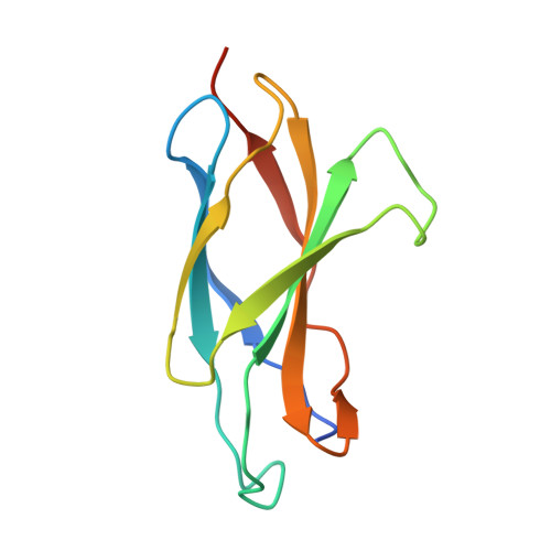
Hedgehog (Hh) signaling proteins stimulate cell proliferation, differentiation, and tissue patterning at multiple points in animal development. A single Hh homolog is present in Drosophila, but three Hh homologs, Sonic Hh, Indian Hh, and Desert Hh, are present in mammals. Distribution, movement, and reception of Hh signals are tightly regulated, and abnormal Hh signaling is associated with developmental defects and cancer. In addition to the integral membrane proteins Patched and Smoothened, members of the Drosophila Ihog family of adhesion-like molecules have recently been shown to bind Hh proteins with micromolar affinity and positively regulate Hh signaling. Cell adhesion molecule-related, down-regulated by oncogenes (CDO) and Brother of CDO (BOC) are the closest mammalian relatives of Drosophila Ihog, and CDO binds Sonic Hh with micromolar affinity and positively regulates Hh signaling. Despite these similarities, structural and biochemical studies have shown that Ihog and CDO utilize nonorthologous domains and completely different binding modes to interact with cognate Hh proteins. We report here biochemical and x-ray structural studies of Sonic, Indian, and Desert Hh proteins both alone and complexed with active domains of CDO and BOC. These results show that all mammalian Hh proteins bind CDO and BOC in the same manner. We also show that interactions between Hh proteins and CDO are weakened at low pH. Formation of Hh-mediated Hh oligomers is thought to be an important feature of normal Hh signaling, but no conserved self-interaction between Hh proteins is apparent from inspection of 14 independent Hh-containing crystal lattices.
- Department of Biophysics and Biophysical Chemistry, Johns Hopkins University School of Medicine, Baltimore, Maryland 21205, USA.
Organizational Affiliation: