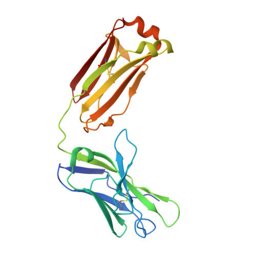
Structure and function of broadly reactive antibody PG16 reveal an H3 subdomain that mediates potent neutralization of HIV-1.
Pejchal, R., Walker, L.M., Stanfield, R.L., Phogat, S.K., Koff, W.C., Poignard, P., Burton, D.R., Wilson, I.A.(2010) Proc Natl Acad Sci U S A 107: 11483-11488
- PubMed: 20534513 Search on PubMedSearch on PubMed Central
- DOI: https://doi.org/10.1073/pnas.1004600107
- Primary Citation Related Structures:
3MUG, 3MUH - PubMed Abstract:
Development of an effective vaccine against HIV-1 will likely require elicitation of broad and potent neutralizing antibodies against the trimeric surface envelope glycoprotein (Env). Monoclonal antibodies (mAbs) PG9 and PG16 neutralize approximately 80% of HIV-1 isolates across all clades with extraordinary potency and target novel epitopes preferentially expressed on Env trimers. As these neutralization properties are ideal for a vaccine-elicited antibody response to HIV-1, their structural basis was investigated. The crystal structure of the antigen-binding fragment (Fab) of PG16 at 2.5 A resolution revealed its unusually long, 28-residue, complementarity determining region (CDR) H3 forms a unique, stable subdomain that towers above the antibody surface. A 7-residue "specificity loop" on the "hammerhead" subdomain was identified that, when transplanted from PG16 to PG9 and vice versa, accounted for differences in the fine specificity and neutralization of these two mAbs. The PG16 electron density maps also revealed that a CDR H3 tyrosine was sulfated, which was confirmed for both PG9 (doubly) and PG16 (singly) by mass spectral analysis. We further showed that tyrosine sulfation plays a role in binding and neutralization. An N-linked glycan modification is observed in the variable light chain, but not required for antigen recognition. Further, the crystal structure of the PG9 light chain at 3.0 A facilitated homology modeling to support the presence of these unusual features in PG9. Thus, PG9 and PG16 use unique structural features to mediate potent neutralization of HIV-1 that may be of utility in antibody engineering and for high-affinity recognition of a variety of therapeutic targets.
- Department of Molecular Biology, Scripps Research Institute, La Jolla, CA 92037, USA.
Organizational Affiliation: