3MPB

Z5688 from E. coli O157:H7 bound to fructose


Experimental Data Snapshot

  • Method: X-RAY DIFFRACTION
  • Resolution: 1.91 Å
  • R-Value Free: 0.233 
  • R-Value Work: 0.188 
  • R-Value Observed: 0.191 

wwPDB Validation   3D Report Full Report


Ligand Structure Quality Assessment 


This is version 1.3 of the entry. See complete history


Literature

Structure-based annotation of a novel sugar isomerase from the pathogenic E. coli O157:H7.

van Staalduinen, L.M.Park, C.S.Yeom, S.J.Adams-Cioaba, M.A.Oh, D.K.Jia, Z.

(2010) J Mol Biol 401: 866-881

  • DOI: https://doi.org/10.1016/j.jmb.2010.06.063
  • Primary Citation of Related Structures:  
    3KMH, 3MPB

  • PubMed Abstract: 

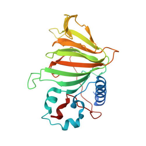
    Prokaryotes can use a variety of sugars as carbon sources in order to provide a selective survival advantage. The gene z5688 found in the pathogenic Escherichia coli O157:H7 encodes a "hypothetical" protein of unknown function. Sequence analysis identified the gene product as a putative member of the cupin superfamily of proteins, but no other functional information was known. We have determined the crystal structure of the Z5688 protein at 1.6 A resolution and identified the protein as a novel E. coli sugar isomerase (EcSI) through overall fold analysis and secondary-structure matching. Extensive substrate screening revealed that EcSI is capable of acting on d-lyxose and d-mannose. The complex structure of EcSI with fructose allowed the identification of key active-site residues, and mutagenesis confirmed their importance. The structure of EcSI also suggested a novel mechanism for substrate binding and product release in a cupin sugar isomerase. Supplementation of a nonpathogenic E. coli strain with EcSI enabled cell growth on the rare pentose d-lyxose.


  • Organizational Affiliation

    Department of Biochemistry, Queen's University, Kingston, Ontario, Canada K7L3N6.


Macromolecules
Find similar proteins by:  (by identity cutoff)  |  3D Structure
Entity ID: 1
MoleculeChains Sequence LengthOrganismDetailsImage
Sugar isomerase
A, B
246Escherichia coli O157:H7Mutation(s): 0 
Gene Names: ECs5070Z5688
UniProt
Find proteins for Q8X5Q7 (Escherichia coli O157:H7)
Explore Q8X5Q7 
Go to UniProtKB:  Q8X5Q7
Entity Groups  
Sequence Clusters30% Identity50% Identity70% Identity90% Identity95% Identity100% Identity
UniProt GroupQ8X5Q7
Sequence Annotations
Expand
  • Reference Sequence
Small Molecules
Ligands 4 Unique
IDChains Name / Formula / InChI Key2D Diagram3D Interactions
FRU
Query on FRU

Download Ideal Coordinates CCD File 
E [auth A],
G [auth B]
beta-D-fructofuranose
C6 H12 O6
RFSUNEUAIZKAJO-ARQDHWQXSA-N
GOL
Query on GOL

Download Ideal Coordinates CCD File 
F [auth A]GLYCEROL
C3 H8 O3
PEDCQBHIVMGVHV-UHFFFAOYSA-N
ACT
Query on ACT

Download Ideal Coordinates CCD File 
D [auth A],
I [auth B]
ACETATE ION
C2 H3 O2
QTBSBXVTEAMEQO-UHFFFAOYSA-M
MN
Query on MN

Download Ideal Coordinates CCD File 
C [auth A],
H [auth B]
MANGANESE (II) ION
Mn
WAEMQWOKJMHJLA-UHFFFAOYSA-N
Experimental Data & Validation

Experimental Data

  • Method: X-RAY DIFFRACTION
  • Resolution: 1.91 Å
  • R-Value Free: 0.233 
  • R-Value Work: 0.188 
  • R-Value Observed: 0.191 
  • Space Group: P 1 21 1
Unit Cell:
Length ( Å )Angle ( ˚ )
a = 53.425α = 90
b = 75.699β = 106.54
c = 62.022γ = 90
Software Package:
Software NamePurpose
Blu-Icedata collection
REFMACrefinement
HKL-2000data reduction
HKL-2000data scaling
REFMACphasing

Structure Validation

View Full Validation Report


Ligand Structure Quality Assessment 


Entry History 

Deposition Data

Revision History  (Full details and data files)

  • Version 1.0: 2010-07-21
    Type: Initial release
  • Version 1.1: 2011-07-13
    Changes: Version format compliance
  • Version 1.2: 2020-07-29
    Type: Remediation
    Reason: Carbohydrate remediation
    Changes: Data collection, Derived calculations, Structure summary
  • Version 1.3: 2023-09-06
    Changes: Data collection, Database references, Refinement description, Structure summary