In vivo protein crystallization opens new routes in structural biology.
Koopmann, R., Cupelli, K., Redecke, L., Nass, K., Deponte, D.P., White, T.A., Stellato, F., Rehders, D., Liang, M., Andreasson, J., Aquila, A., Bajt, S., Barthelmess, M., Barty, A., Bogan, M.J., Bostedt, C., Boutet, S., Bozek, J.D., Caleman, C., Coppola, N., Davidsson, J., Doak, R.B., Ekeberg, T., Epp, S.W., Erk, B., Fleckenstein, H., Foucar, L., Graafsma, H., Gumprecht, L., Hajdu, J., Hampton, C.Y., Hartmann, A., Hartmann, R., Hauser, G., Hirsemann, H., Holl, P., Hunter, M.S., Kassemeyer, S., Kirian, R.A., Lomb, L., Maia, F.R., Kimmel, N., Martin, A.V., Messerschmidt, M., Reich, C., Rolles, D., Rudek, B., Rudenko, A., Schlichting, I., Schulz, J., Seibert, M.M., Shoeman, R.L., Sierra, R.G., Soltau, H., Stern, S., Struder, L., Timneanu, N., Ullrich, J., Wang, X., Weidenspointner, G., Weierstall, U., Williams, G.J., Wunderer, C.B., Fromme, P., Spence, J.C., Stehle, T., Chapman, H.N., Betzel, C., Duszenko, M.(2012) Nat Methods 9: 259-262
- PubMed: 22286384 Search on PubMedSearch on PubMed Central
- DOI: https://doi.org/10.1038/nmeth.1859
- Primary Citation Related Structures:
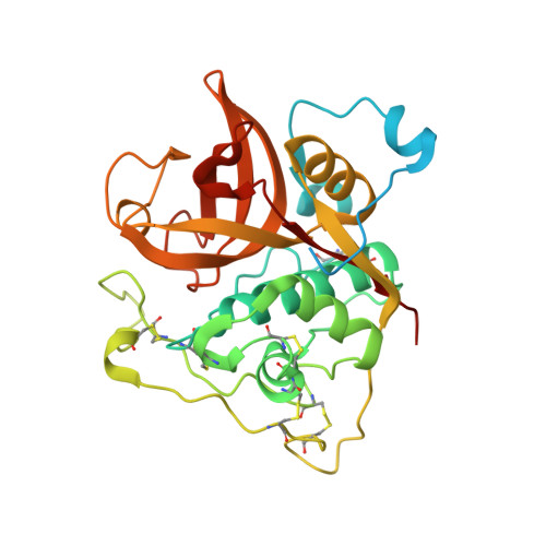
3MOR - PubMed Abstract:
Protein crystallization in cells has been observed several times in nature. However, owing to their small size these crystals have not yet been used for X-ray crystallographic analysis. We prepared nano-sized in vivo-grown crystals of Trypanosoma brucei enzymes and applied the emerging method of free-electron laser-based serial femtosecond crystallography to record interpretable diffraction data. This combined approach will open new opportunities in structural systems biology.
- Interfaculty Institute of Biochemistry, University of Tübingen, Tübingen, Germany.
Organizational Affiliation: