3MN8

Structure of Drosophila melanogaster carboxypeptidase D isoform 1B short


Experimental Data Snapshot

  • Method: X-RAY DIFFRACTION
  • Resolution: 2.70 Å
  • R-Value Free: 0.282 
  • R-Value Work: 0.211 
  • R-Value Observed: 0.212 

wwPDB Validation   3D Report Full Report


Ligand Structure Quality Assessment 


This is version 1.4 of the entry. See complete history


Literature

Structure-Function Analysis of the Short Splicing Variant Carboxypeptidase Encoded by Drosophila melanogaster silver.

Tanco, S.Arolas, J.L.Guevara, T.Lorenzo, J.Aviles, F.X.Gomis-Ruth, F.X.

(2010) J Mol Biol 401: 465-477

  • DOI: https://doi.org/10.1016/j.jmb.2010.06.035
  • Primary Citation of Related Structures:  
    3MN8

  • PubMed Abstract: 

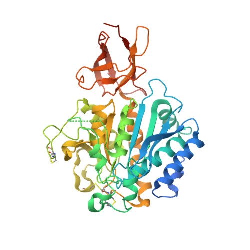
    Drosophila melanogaster silver gene is the ortholog of the coding gene of mammalian carboxypeptidase D (CPD). The silver gene gives rise to eight different splicing variants of differing length that can contain up to three homologous repeats. Among the protein variants encoded, the short form 1B alias DmCPD1Bs (D. melanogaster CPD variant 1B short) is necessary and sufficient for viability of the fruit fly. It has one single repeat, it is active against standard peptide substrates, and it is localized to the secretory pathway. In this work, the enzyme was found as a monomer in solution and as a homodimer in the crystal structure, which features a protomer with an N-terminal 311-residue catalytic domain of alpha/beta-hydrolase fold and a C-terminal 84-residue all-beta transthyretin-like domain. Overall, DmCPD1Bs conforms to the structure of N/E-type funnelins/M14B metallopeptidases, but it has two unique structural elements potentially involved in regulation of its activity: (i) two contiguous surface cysteines that may become palmitoylated and target the enzyme to membranes, thus providing control through localization, and (ii) a surface hot spot targetable by peptidases that would provide a regulatory mechanism through proteolytic inactivation. Given that the fruit fly possesses orthologs of only two out of the five proteolytically competent N/E-type funnelins found in higher vertebrates, DmCPD1Bs may represent a functional analog of at least one of the missing mammalian CPs.


  • Organizational Affiliation

    Departament de Bioquímica i Biologia Molecular, Institut de Biotecnologia i de Biomedicina, Universitat Autònoma de Barcelona, E-08193 Bellaterra, Spain.


Macromolecules
Find similar proteins by:  (by identity cutoff)  |  3D Structure
Entity ID: 1
MoleculeChains Sequence LengthOrganismDetailsImage
LP15968p
A, B, C, D
435Drosophila melanogasterMutation(s): 0 
Gene Names: svr-RF
UniProt
Find proteins for P42787 (Drosophila melanogaster)
Explore P42787 
Go to UniProtKB:  P42787
Entity Groups  
Sequence Clusters30% Identity50% Identity70% Identity90% Identity95% Identity100% Identity
UniProt GroupP42787
Sequence Annotations
Expand
  • Reference Sequence
Small Molecules
Ligands 4 Unique
IDChains Name / Formula / InChI Key2D Diagram3D Interactions
GEM
Query on GEM

Download Ideal Coordinates CCD File 
G [auth A],
M [auth B],
P [auth C],
T [auth D]
(2-GUANIDINOETHYLMERCAPTO)SUCCINIC ACID
C7 H13 N3 O4 S
VKVCLXDFOQQABP-BYPYZUCNSA-N
NAG
Query on NAG

Download Ideal Coordinates CCD File 
E [auth A],
K [auth B],
N [auth C],
R [auth D]
2-acetamido-2-deoxy-beta-D-glucopyranose
C8 H15 N O6
OVRNDRQMDRJTHS-FMDGEEDCSA-N
GOL
Query on GOL

Download Ideal Coordinates CCD File 
H [auth A],
I [auth A],
J [auth A],
Q [auth C]
GLYCEROL
C3 H8 O3
PEDCQBHIVMGVHV-UHFFFAOYSA-N
ZN
Query on ZN

Download Ideal Coordinates CCD File 
F [auth A],
L [auth B],
O [auth C],
S [auth D]
ZINC ION
Zn
PTFCDOFLOPIGGS-UHFFFAOYSA-N
Binding Affinity Annotations 
IDSourceBinding Affinity
GEM PDBBind:  3MN8 Ki: 670 (nM) from 1 assay(s)
Experimental Data & Validation

Experimental Data

  • Method: X-RAY DIFFRACTION
  • Resolution: 2.70 Å
  • R-Value Free: 0.282 
  • R-Value Work: 0.211 
  • R-Value Observed: 0.212 
  • Space Group: P 21 21 21
Unit Cell:
Length ( Å )Angle ( ˚ )
a = 97.02α = 90
b = 135.7β = 90
c = 141.71γ = 90
Software Package:
Software NamePurpose
ProDCdata collection
PHASERphasing
PHENIXrefinement
XDSdata reduction
SCALAdata scaling

Structure Validation

View Full Validation Report


Ligand Structure Quality Assessment 


Entry History 

Deposition Data

Revision History  (Full details and data files)

  • Version 1.0: 2010-07-28
    Type: Initial release
  • Version 1.1: 2011-07-13
    Changes: Version format compliance
  • Version 1.2: 2017-11-08
    Changes: Refinement description
  • Version 1.3: 2020-07-29
    Type: Remediation
    Reason: Carbohydrate remediation
    Changes: Data collection, Derived calculations, Structure summary
  • Version 1.4: 2023-09-06
    Changes: Data collection, Database references, Refinement description, Structure summary