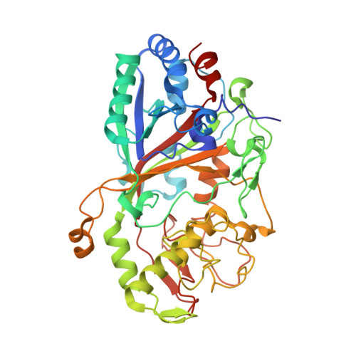
3MM3

Dye-decolorizing peroxidase (DyP) D171N in complex with cyanide


Experimental Data Snapshot

  • Method: X-RAY DIFFRACTION
  • Resolution: 1.40 Å
  • R-Value Free: 0.189 
  • R-Value Work: 0.181 
  • R-Value Observed: 0.182 

Starting Model: experimental
View more details

wwPDB Validation   3D Report Full Report


Ligand Structure Quality Assessment 


This is version 1.4 of the entry. See complete history


Literature

The catalytic mechanism of dye-decolorizing peroxidase DyP may require the swinging movement of an aspartic acid residue

Yoshida, T.Tsuge, H.Konno, H.Hisabori, T.Sugano, Y.

(2011) FEBS J 278: 2387-2394

  • DOI: https://doi.org/10.1111/j.1742-4658.2011.08161.x
  • Primary Citation of Related Structures:  
    2D3Q, 3AFV, 3MM1, 3MM2, 3MM3

  • PubMed Abstract: 

    The dye-decolorizing peroxidase (DyP)-type peroxidase family is a unique heme peroxidase family. The primary and tertiary structures of this family are obviously different from those of other heme peroxidases. However, the details of the structure-function relationships of this family remain poorly understood. We show four high-resolution structures of DyP (EC1.11.1.19), which is representative of this family: the native DyP (1.40 Å), the D171N mutant DyP (1.42 Å), the native DyP complexed with cyanide (1.45 Å), and the D171N mutant DyP associated with cyanide (1.40 Å). These structures contain four amino acids forming the binding pocket for hydrogen peroxide, and they are remarkably conserved in this family. Moreover, these structures show that OD2 of Asp171 accepts a proton from hydrogen peroxide in compound I formation, and that OD2 can swing to the appropriate position in response to the ligand for heme iron. On the basis of these results, we propose a swing mechanism in compound I formation. When DyP reacts with hydrogen peroxide, OD2 swings towards an optimal position to accept the proton from hydrogen peroxide bound to the heme iron.


  • Organizational Affiliation

    R1-7 Chemical Resources Laboratory, Tokyo Institute of Technology, Japan.


Macromolecules
Find similar proteins by:  (by identity cutoff)  |  3D Structure
Entity ID: 1
MoleculeChains Sequence LengthOrganismDetailsImage
DyP442Bjerkandera adustaMutation(s): 1 
Gene Names: dyp
EC: 1.11.1.19
UniProt
Find proteins for Q8WZK8 (Bjerkandera adusta)
Explore Q8WZK8 
Go to UniProtKB:  Q8WZK8
Entity Groups  
Sequence Clusters30% Identity50% Identity70% Identity90% Identity95% Identity100% Identity
UniProt GroupQ8WZK8
Sequence Annotations
Expand
  • Reference Sequence
Small Molecules
Ligands 2 Unique
IDChains Name / Formula / InChI Key2D Diagram3D Interactions
HEM
Query on HEM

Download Ideal Coordinates CCD File 
B [auth A]PROTOPORPHYRIN IX CONTAINING FE
C34 H32 Fe N4 O4
KABFMIBPWCXCRK-RGGAHWMASA-L
CYN
Query on CYN

Download Ideal Coordinates CCD File 
C [auth A]CYANIDE ION
C N
XFXPMWWXUTWYJX-UHFFFAOYSA-N
Experimental Data & Validation

Experimental Data

  • Method: X-RAY DIFFRACTION
  • Resolution: 1.40 Å
  • R-Value Free: 0.189 
  • R-Value Work: 0.181 
  • R-Value Observed: 0.182 
  • Space Group: P 1 21 1
Unit Cell:
Length ( Å )Angle ( ˚ )
a = 44.954α = 90
b = 97.15β = 104.04
c = 46.46γ = 90
Software Package:
Software NamePurpose
DENZOdata reduction
SCALEPACKdata scaling
MOLREPphasing
REFMACrefinement
PDB_EXTRACTdata extraction

Structure Validation

View Full Validation Report


Ligand Structure Quality Assessment 


Entry History 

Deposition Data

Revision History  (Full details and data files)

  • Version 1.0: 2011-04-27
    Type: Initial release
  • Version 1.1: 2011-07-13
    Changes: Version format compliance
  • Version 1.2: 2011-10-26
    Changes: Database references, Source and taxonomy
  • Version 1.3: 2017-11-08
    Changes: Refinement description
  • Version 1.4: 2023-11-01
    Changes: Data collection, Database references, Derived calculations, Refinement description