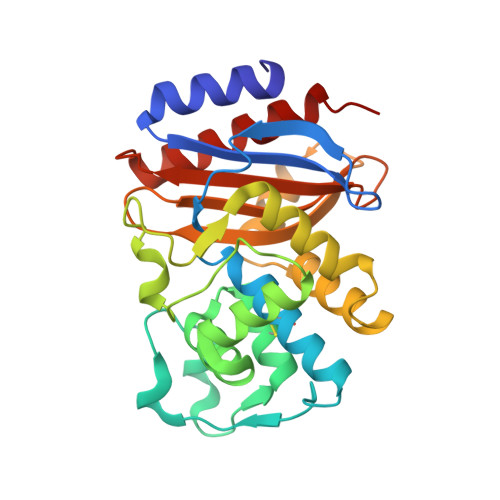
3MKE

SHV-1 beta-lactamase complex with LP06


Experimental Data Snapshot

  • Method: X-RAY DIFFRACTION
  • Resolution: 1.75 Å
  • R-Value Free: 0.181 
  • R-Value Work: 0.153 
  • R-Value Observed: 0.155 

Starting Model: experimental
View more details

wwPDB Validation   3D Report Full Report


Ligand Structure Quality Assessment 


This is version 1.5 of the entry. See complete history


Literature

Novel Insights into the Mode of Inhibition of Class A SHV-1 {beta}-Lactamases Revealed by Boronic Acid Transition State Inhibitors.

Ke, W.Sampson, J.M.Ori, C.Prati, F.Drawz, S.M.Bethel, C.R.Bonomo, R.A.van den Akker, F.

(2011) Antimicrob Agents Chemother 55: 174-183

  • DOI: https://doi.org/10.1128/AAC.00930-10
  • Primary Citation of Related Structures:  
    3MKE, 3MKF, 3MXR, 3MXS

  • PubMed Abstract: 

    Boronic acid transition state inhibitors (BATSIs) are potent class A and C β-lactamase inactivators and are of particular interest due to their reversible nature mimicking the transition state. Here, we present structural and kinetic data describing the inhibition of the SHV-1 β-lactamase, a clinically important enzyme found in Klebsiella pneumoniae, by BATSI compounds possessing the R1 side chains of ceftazidime and cefoperazone and designed variants of the latter, compounds 1 and 2. The ceftazidime and cefoperazone BATSI compounds inhibit the SHV-1 β-lactamase with micromolar affinity that is considerably weaker than their inhibition of other β-lactamases. The solved crystal structures of these two BATSIs in complex with SHV-1 reveal a possible reason for SHV-1's relative resistance to inhibition, as the BATSIs adopt a deacylation transition state conformation compared to the usual acylation transition state conformation when complexed to other β-lactamases. Active-site comparison suggests that these conformational differences might be attributed to a subtle shift of residue A237 in SHV-1. The ceftazidime BATSI structure revealed that the carboxyl-dimethyl moiety is positioned in SHV-1's carboxyl binding pocket. In contrast, the cefoperazone BATSI has its R1 group pointing away from the active site such that its phenol moiety moves residue Y105 from the active site via end-on stacking interactions. To work toward improving the affinity of the cefoperazone BATSI, we synthesized two variants in which either one or two extra carbons were added to the phenol linker. Both variants yielded improved affinity against SHV-1, possibly as a consequence of releasing the strain of its interaction with the unusual Y105 conformation.


  • Organizational Affiliation

    Department of Biochemistry, Case Western Reserve University, Cleveland, OH 44106-4935, USA.


Macromolecules
Find similar proteins by:  (by identity cutoff)  |  3D Structure
Entity ID: 1
MoleculeChains Sequence LengthOrganismDetailsImage
Beta-lactamase SHV-1265Klebsiella pneumoniaeMutation(s): 0 
Gene Names: blashv1
EC: 3.5.2.6
UniProt
Find proteins for P0AD64 (Klebsiella pneumoniae)
Explore P0AD64 
Go to UniProtKB:  P0AD64
Entity Groups  
Sequence Clusters30% Identity50% Identity70% Identity90% Identity95% Identity100% Identity
UniProt GroupP0AD64
Sequence Annotations
Expand
  • Reference Sequence
Small Molecules
Ligands 3 Unique
IDChains Name / Formula / InChI Key2D Diagram3D Interactions
MA4
Query on MA4

Download Ideal Coordinates CCD File 
B [auth A],
C [auth A]
CYCLOHEXYL-HEXYL-BETA-D-MALTOSIDE
C24 H44 O11
WUCWJXGMSXTDAV-QKMCSOCLSA-N
CZ6
Query on CZ6

Download Ideal Coordinates CCD File 
E [auth A]2-[(~{Z})-[1-(2-azanyl-1,3-thiazol-4-yl)-2-oxidanylidene-2-[[(6~{S})-4,4,6-trimethyl-1,3,2-dioxaborinan-2-yl]methylamino]ethylidene]amino]oxy-2-methyl-propanoic acid
C16 H25 B N4 O6 S
VUZFEPQBACFWNR-GAGKSFBISA-N
CB4
Query on CB4

Download Ideal Coordinates CCD File 
D [auth A]PINACOL[[2-AMINO-ALPHA-(1-CARBOXY-1-METHYLETHOXYIMINO)-4-THIAZOLEACETYL]AMINO]METHANEBORONATE
C10 H15 B N4 O6 S
ZECCQELUYUPTSB-UUASQNMZSA-N
Binding Affinity Annotations 
IDSourceBinding Affinity
CB4 PDBBind:  3MKE Ki: 2200 (nM) from 1 assay(s)
Experimental Data & Validation

Experimental Data

  • Method: X-RAY DIFFRACTION
  • Resolution: 1.75 Å
  • R-Value Free: 0.181 
  • R-Value Work: 0.153 
  • R-Value Observed: 0.155 
  • Space Group: P 21 21 21
Unit Cell:
Length ( Å )Angle ( ˚ )
a = 49.554α = 90
b = 56.484β = 90
c = 82.067γ = 90
Software Package:
Software NamePurpose
HKL-2000data collection
REFMACrefinement
HKL-2000data reduction
HKL-2000data scaling

Structure Validation

View Full Validation Report


Ligand Structure Quality Assessment 


Entry History 

Deposition Data

Revision History  (Full details and data files)

  • Version 1.0: 2010-11-24
    Type: Initial release
  • Version 1.1: 2011-07-13
    Changes: Version format compliance
  • Version 1.2: 2017-11-08
    Changes: Refinement description
  • Version 1.3: 2020-02-19
    Changes: Derived calculations, Structure summary
  • Version 1.4: 2023-09-06
    Changes: Data collection, Database references, Derived calculations, Refinement description
  • Version 1.5: 2024-11-06
    Changes: Structure summary