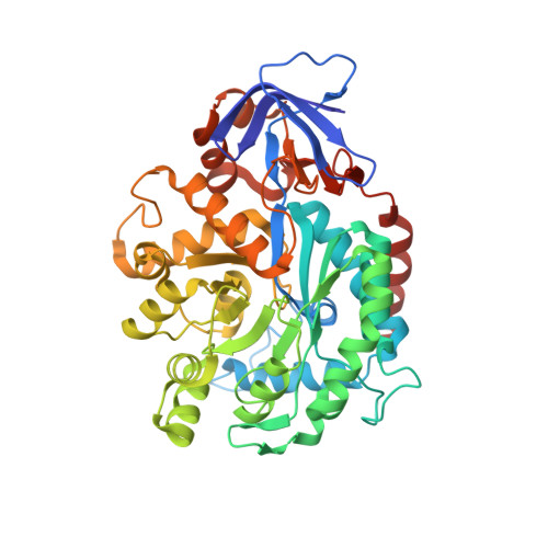
3MDW

The structure of N-formimino-L-Glutamate Iminohydrolase from Pseudomonas aeruginosa complexed with N-formimino-L-Aspartate


Experimental Data Snapshot

  • Method: X-RAY DIFFRACTION
  • Resolution: 1.90 Å
  • R-Value Free: 0.203 
  • R-Value Work: 0.170 
  • R-Value Observed: 0.171 

wwPDB Validation   3D Report Full Report


Ligand Structure Quality Assessment 


This is version 1.5 of the entry. See complete history


Literature

Structure of N-Formimino-l-glutamate Iminohydrolase from Pseudomonas aeruginosa.

Fedorov, A.A.Marti-Arbona, R.Nemmara, V.V.Hitchcock, D.Fedorov, E.V.Almo, S.C.Raushel, F.M.

(2015) Biochemistry 54: 890-897

  • DOI: https://doi.org/10.1021/bi501299y
  • Primary Citation of Related Structures:  
    3MDU, 3MDW, 4RZB

  • PubMed Abstract: 

    N-Formimino-l-glutamate iminohydrolase (HutF), from Pseudomonas aeruginosa with a locus tag of Pa5106 ( gi|15600299 ), is a member of the amidohydrolase superfamily. This enzyme catalyzes the deamination of N-formimino-l-glutamate to N-formyl-l-glutamate and ammonia in the histidine degradation pathway. The crystal structure of Pa5106 was determined in the presence of the inhibitors N-formimino-l-aspartate and N-guanidino-l-glutaric acid at resolutions of 1.9 and 1.4 Å, respectively. The structure of an individual subunit is composed of two domains with the larger domain folding as a distorted (β/α)8-barrel. The (β/α)8-barrel domain is composed of eight β-strands flanked by 11 α-helices, whereas the smaller domain is made up of eight β-strands. The active site of Pa5106 contains a single zinc atom that is coordinated by His-56, His-58, His-232, and Asp-320. The nucleophilic solvent water molecule coordinates with the zinc atom at a distance of 2.0 Å and is hydrogen bonded to Asp-320 and His-269. The α-carboxylate groups of both inhibitors are hydrogen bonded to the imidazole moiety of His-206, the hydroxyl group of Tyr-121, and the side chain amide group of Gln-61. The side chain carboxylate groups of the two inhibitors are ion-paired with the guanidino groups of Arg-209 and Arg-82. Computational docking of high-energy tetrahedral intermediate forms of the substrate, N-formimino-l-glutamate, to the three-dimensional structure of Pa5106 suggests that this compound likely undergoes a re-faced nucleophilic attack at the formimino group by the metal-bound hydroxide. A catalytic mechanism of the reaction catalyzed by Pa5106 is proposed.


  • Organizational Affiliation

    Department of Chemistry and ‡Department of Biochemistry & Biophysics, Texas A&M University , College Station, Texas 77843, United States.


Macromolecules
Find similar proteins by:  (by identity cutoff)  |  3D Structure
Entity ID: 1
MoleculeChains Sequence LengthOrganismDetailsImage
N-formimino-L-Glutamate Iminohydrolase
A, B, C, D
453Pseudomonas aeruginosaMutation(s): 0 
Gene Names: PA5106
UniProt
Find proteins for Q9HU77 (Pseudomonas aeruginosa (strain ATCC 15692 / DSM 22644 / CIP 104116 / JCM 14847 / LMG 12228 / 1C / PRS 101 / PAO1))
Explore Q9HU77 
Go to UniProtKB:  Q9HU77
Entity Groups  
Sequence Clusters30% Identity50% Identity70% Identity90% Identity95% Identity100% Identity
UniProt GroupQ9HU77
Sequence Annotations
Expand
  • Reference Sequence
Small Molecules
Ligands 4 Unique
IDChains Name / Formula / InChI Key2D Diagram3D Interactions
NFQ
Query on NFQ

Download Ideal Coordinates CCD File 
E [auth A],
H [auth B],
J [auth C],
M [auth D]
N-[(E)-iminomethyl]-L-aspartic acid
C5 H8 N2 O4
XTPIFIMCFHNJOH-VKHMYHEASA-N
PO4
Query on PO4

Download Ideal Coordinates CCD File 
O [auth D]PHOSPHATE ION
O4 P
NBIIXXVUZAFLBC-UHFFFAOYSA-K
GOL
Query on GOL

Download Ideal Coordinates CCD File 
G [auth A],
L [auth C]
GLYCEROL
C3 H8 O3
PEDCQBHIVMGVHV-UHFFFAOYSA-N
ZN
Query on ZN

Download Ideal Coordinates CCD File 
F [auth A],
I [auth B],
K [auth C],
N [auth D]
ZINC ION
Zn
PTFCDOFLOPIGGS-UHFFFAOYSA-N
Experimental Data & Validation

Experimental Data

  • Method: X-RAY DIFFRACTION
  • Resolution: 1.90 Å
  • R-Value Free: 0.203 
  • R-Value Work: 0.170 
  • R-Value Observed: 0.171 
  • Space Group: C 1 2 1
Unit Cell:
Length ( Å )Angle ( ˚ )
a = 304.046α = 90
b = 67.247β = 91.5
c = 98.229γ = 90
Software Package:
Software NamePurpose
ADSCdata collection
PHENIXmodel building
PHENIXrefinement
DENZOdata reduction
SCALEPACKdata scaling
PHENIXphasing

Structure Validation

View Full Validation Report


Ligand Structure Quality Assessment 


Entry History 

Deposition Data

Revision History  (Full details and data files)

  • Version 1.0: 2011-03-09
    Type: Initial release
  • Version 1.1: 2011-07-13
    Changes: Version format compliance
  • Version 1.2: 2015-01-21
    Changes: Database references
  • Version 1.3: 2015-01-28
    Changes: Database references
  • Version 1.4: 2015-02-11
    Changes: Database references
  • Version 1.5: 2024-02-21
    Changes: Data collection, Database references, Derived calculations