3M7Q | pdb_00003m7q

Crystal structure of recombinant Kunitz Type serine protease Inhibitor-1 from the Caribbean sea anemone stichodactyla helianthus in complex with bovine pancreatic trypsin


Experimental Data Snapshot

  • Method: X-RAY DIFFRACTION
  • Resolution: 1.70 Å
  • R-Value Free: 
    0.188 (Depositor), 0.194 (DCC) 
  • R-Value Work: 
    0.158 (Depositor) 
  • R-Value Observed: 
    0.159 (Depositor) 

Starting Model: experimental
View more details

wwPDB Validation 3D Report Full Report

Validation slider image for 3M7Q

This is version 1.5 of the entry. See complete history

Literature

Structural insights into serine protease inhibition by a marine invertebrate BPTI Kunitz-type inhibitor.

Garcia-Fernandez, R.Pons, T.Perbandt, M.Valiente, P.A.Talavera, A.Gonzalez-Gonzalez, Y.Rehders, D.Chavez, M.A.Betzel, C.Redecke, L.

(2012) J Struct Biol 180: 271-279

  • DOI: https://doi.org/10.1016/j.jsb.2012.08.009
  • Primary Citation Related Structures: 
    3M7Q

  • PubMed Abstract: 

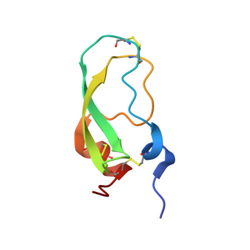
    Proteins isolated from marine invertebrates are frequently characterized by exceptional structural and functional properties. ShPI-1, a BPTI Kunitz-type inhibitor from the Caribbean Sea anemone Stichodactyla helianthus, displays activity not only against serine-, but also against cysteine-, and aspartate proteases. As an initial step to evaluate the molecular basis of its activities, we describe the crystallographic structure of ShPI-1 in complex with the serine protease bovine pancreatic trypsin at 1.7Å resolution. The overall structure and the important enzyme-inhibitor interactions of this first invertebrate BPTI-like Kunitz-type inhibitor:trypsin complex remained largely conserved compared to mammalian BPTI-Kunitz inhibitor complexes. However, a prominent stabilizing role within the interface was attributed to arginine at position P3. Binding free-energy calculations indicated a 10-fold decrease for the inhibitor affinity against trypsin, if the P3 residue of ShPI-1 is mutated to alanine. Together with the increased role of Arg(11) at P3 position, slightly reduced interactions at the prime side (Pn') of the primary binding loop and at the secondary binding loop of ShPI-1 were detected. In addition, the structure provides important information for site directed mutagenesis to further optimize the activity of rShPI-1A for biotechnological applications.


  • Organizational Affiliation
    • Centro de Estudio de Proteínas, Facultad de Biología, Universidad de la Habana, Calle 25 No 411, Havana, Cuba.

Macromolecule Content 

  • Total Structure Weight: 30.11 kDa 
  • Atom Count: 2,698 
  • Modeled Residue Count: 284 
  • Deposited Residue Count: 284 
  • Unique protein chains: 2

Macromolecules

Find similar proteins by:|  3D Structure
Entity ID: 1
MoleculeChains  Sequence LengthOrganismDetailsImage
Cationic trypsin223Bos taurusMutation(s): 0 
EC: 3.4.21.4
UniProt
Find proteins for P00760 (Bos taurus)
Explore P00760 
Go to UniProtKB:  P00760
Entity Groups
Sequence Clusters30% Identity50% Identity70% Identity90% Identity95% Identity100% Identity
UniProt GroupP00760
Sequence Annotations
Expand
Reference Sequence
Find similar proteins by:|  3D Structure
Entity ID: 2
MoleculeChains  Sequence LengthOrganismDetailsImage
Kunitz-type proteinase inhibitor SHPI-161Stichodactyla helianthusMutation(s): 0 
UniProt
Find proteins for P31713 (Stichodactyla helianthus)
Explore P31713 
Go to UniProtKB:  P31713
Entity Groups
Sequence Clusters30% Identity50% Identity70% Identity90% Identity95% Identity100% Identity
UniProt GroupP31713
Sequence Annotations
Expand
Reference Sequence

Small Molecules

Ligands 1 Unique
IDChains Name / Formula / InChI Key2D Diagram3D Interactions
PO4

Query on PO4


Download:Ideal Coordinates CCD File
C [auth A]PHOSPHATE ION
O4 P
NBIIXXVUZAFLBC-UHFFFAOYSA-K

Experimental Data & Validation

Experimental Data

  • Method: X-RAY DIFFRACTION
  • Resolution: 1.70 Å
  • R-Value Free:  0.188 (Depositor), 0.194 (DCC) 
  • R-Value Work:  0.158 (Depositor) 
  • R-Value Observed: 0.159 (Depositor) 
Space Group: P 21 21 21
Unit Cell:
Length ( Å )Angle ( ˚ )
a = 58.279α = 90
b = 66.551β = 90
c = 70.937γ = 90
Software Package:
Software NamePurpose
REFMACrefinement

Structure Validation

View Full Validation Report


Entry History 

Deposition Data

Revision History  (Full details and data files)

  • Version 1.0: 2011-03-16
    Type: Initial release
  • Version 1.1: 2011-07-13
    Changes: Version format compliance
  • Version 1.2: 2012-10-03
    Changes: Database references
  • Version 1.3: 2012-11-14
    Changes: Database references
  • Version 1.4: 2023-09-06
    Changes: Data collection, Database references, Derived calculations, Refinement description
  • Version 1.5: 2024-11-27
    Changes: Structure summary