3M1L

Crystal structure of a C-terminal trunacted mutant of a putative ketoacyl reductase (FabG4) from Mycobacterium tuberculosis H37Rv at 2.5 Angstrom resolution


Experimental Data Snapshot

  • Method: X-RAY DIFFRACTION
  • Resolution: 2.52 Å
  • R-Value Free: 0.241 
  • R-Value Work: 0.181 
  • R-Value Observed: 0.183 

wwPDB Validation   3D Report Full Report


This is version 1.4 of the entry. See complete history


Literature

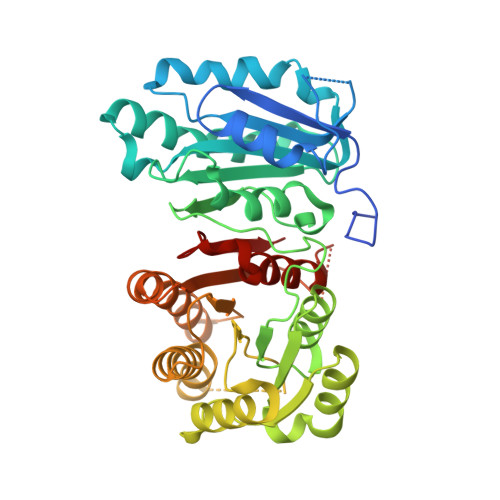
Crystal structure of FabG4 from Mycobacterium tuberculosis reveals the importance of C-terminal residues in ketoreductase activity

Dutta, D.Bhattacharyya, S.Mukherjee, S.Saha, B.Das, A.K.

(2011) J Struct Biol 174: 147-155

  • DOI: https://doi.org/10.1016/j.jsb.2010.11.012
  • Primary Citation of Related Structures:  
    3M1L

  • PubMed Abstract: 

    Rv0242c, also known as FabG4, is a beta-ketoacyl CoA reductase in Mycobacterium tuberculosis. The crystal structure of C-terminal truncated FabG4 is solved at 2.5Å resolution which shows the presence of two distinct domains, domain I and II. Domain I partially resembles "flavodoxin type domain" and the domain II is a typical "ketoacyl CoA reductase (KAR) domain". The enzyme exhibits ketoacyl CoA reductase activity by reducing acetoacyl CoA to 3-hydroxyacyl CoA in presence of NADH. Conserved catalytic triad Ser347, Tyr360, and Lys364 constitute the active site residues of the KAR domain. Presence of the Tyr and the Lys residues in the triad in a particular orientation is imperative for effective catalytic mechanism. The importance of loop I and II and the role of the C-terminal residues of KAR domain are highlighted. Comparative structural analyses clearly demonstrate that loop II is stabilized by hydrophobic interaction with C-terminal residues to sustain the orientation of Tyr360. Loop I interacts with loop II via H-bonding network to restrict the active site residue Lys364 in a catalytically favorable orientation.


  • Organizational Affiliation

    Department of Biotechnology, Indian Institute of Technology Kharagpur, Kharagpur 721032, West Bengal, India.


Macromolecules
Find similar proteins by:  (by identity cutoff)  |  3D Structure
Entity ID: 1
MoleculeChains Sequence LengthOrganismDetailsImage
3-oxoacyl-(Acyl-carrier-protein) reductase
A, B
432Mycobacterium tuberculosis H37RvMutation(s): 0 
Gene Names: Rv0242c
EC: 1.1.1.100
UniProt
Find proteins for O53665 (Mycobacterium tuberculosis (strain CDC 1551 / Oshkosh))
Explore O53665 
Go to UniProtKB:  O53665
Entity Groups  
Sequence Clusters30% Identity50% Identity70% Identity90% Identity95% Identity100% Identity
UniProt GroupO53665
Sequence Annotations
Expand
  • Reference Sequence
Small Molecules
Ligands 1 Unique
IDChains Name / Formula / InChI Key2D Diagram3D Interactions
ACT
Query on ACT

Download Ideal Coordinates CCD File 
C [auth A]ACETATE ION
C2 H3 O2
QTBSBXVTEAMEQO-UHFFFAOYSA-M
Experimental Data & Validation

Experimental Data

  • Method: X-RAY DIFFRACTION
  • Resolution: 2.52 Å
  • R-Value Free: 0.241 
  • R-Value Work: 0.181 
  • R-Value Observed: 0.183 
  • Space Group: P 21 21 21
Unit Cell:
Length ( Å )Angle ( ˚ )
a = 65.076α = 90
b = 80.497β = 90
c = 153.953γ = 90
Software Package:
Software NamePurpose
REFMACrefinement
PDB_EXTRACTdata extraction
StructureStudiodata collection
XDSdata reduction
SCALAdata scaling
PHASERphasing

Structure Validation

View Full Validation Report


Entry History 

Deposition Data

Revision History  (Full details and data files)

  • Version 1.0: 2010-12-22
    Type: Initial release
  • Version 1.1: 2011-07-13
    Changes: Version format compliance
  • Version 1.2: 2014-02-12
    Changes: Database references
  • Version 1.3: 2020-09-16
    Changes: Derived calculations, Structure summary
  • Version 1.4: 2024-03-20
    Changes: Data collection, Database references