3LWR | pdb_00003lwr

Structure of H/ACA RNP bound to a substrate RNA containing 4SU


Experimental Data Snapshot

  • Method: X-RAY DIFFRACTION
  • Resolution: 2.20 Å
  • R-Value Free: 
    0.259 (Depositor), 0.255 (DCC) 
  • R-Value Work: 
    0.222 (Depositor), 0.218 (DCC) 
  • R-Value Observed: 
    0.223 (Depositor) 

wwPDB Validation 3D Report Full Report

Validation slider image for 3LWR

This is version 1.2 of the entry. See complete history

Literature

Functional and Structural Impact of Target Uridine Substitutions on the H/ACA Ribonucleoprotein Particle Pseudouridine Synthase .

Zhou, J.Liang, B.Li, H.

(2010) Biochemistry 49: 6276-6281

  • DOI: https://doi.org/10.1021/bi1006699
  • Primary Citation Related Structures: 
    3LWQ, 3LWR, 3LWV

  • PubMed Abstract: 

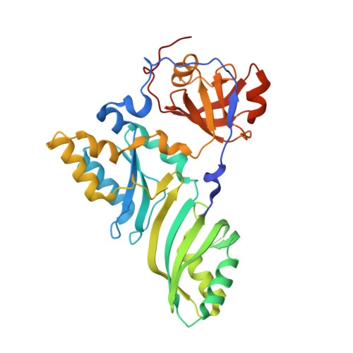
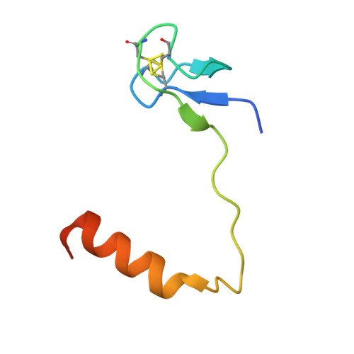
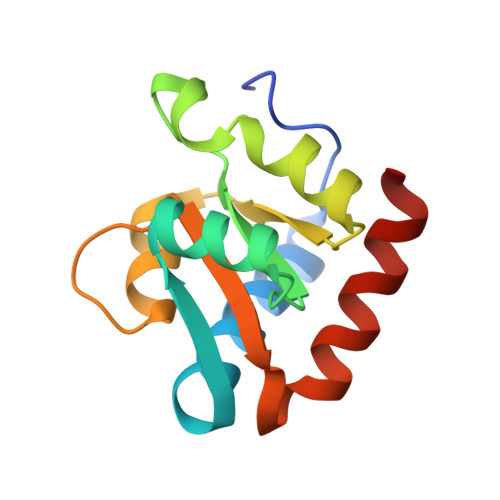
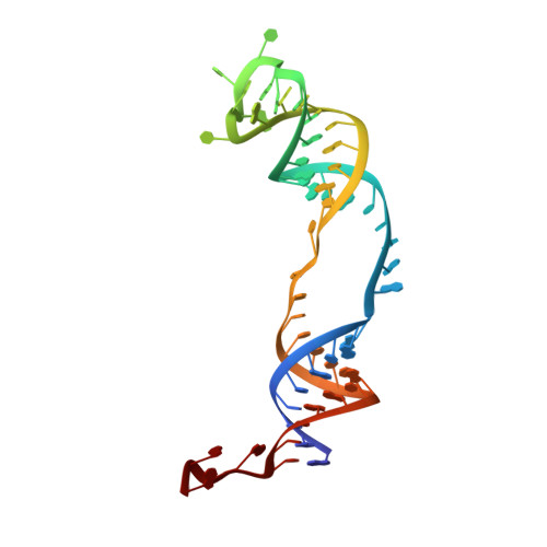
    Box H/ACA ribonucleoprotein protein particles catalyze the majority of pseudouridylation in functional RNA. Different from stand alone pseudouridine synthases, the RNP pseudouridine synthase comprises multiple protein subunits and an RNA subunit. Previous studies showed that each subunit, regardless its location, is sensitive to the step of subunit placement at the catalytic center and potentially to the reaction status of the substrate. Here we describe the impact of chemical substitutions of target uridine on enzyme activity and structure. We found that 3-methyluridine in place of uridine inhibited its isomerization while 2'-deoxyuridine or 4-thiouridine did not. Significantly, crystal structures of an archaeal box H/ACA RNP bound with the nonreactive and the two postreactive substrate analogues showed only subtle structural changes throughout the assembly except for a conserved tyrosine and a substrate anchoring loop of Cbf5. Our results suggest a potential role of these elements and the subunit that contacts them in substrate binding and product release.


  • Organizational Affiliation
    • Department of Chemistry and Biochemistry, Florida State University, Tallahassee, Florida 32306, USA.

Macromolecule Content 

  • Total Structure Weight: 82.24 kDa 
  • Atom Count: 5,432 
  • Modeled Residue Count: 560 
  • Deposited Residue Count: 594 
  • Unique protein chains: 3
  • Unique nucleic acid chains: 2

Macromolecules


Find similar proteins by:|  3D Structure
Entity ID: 1
MoleculeChains  Sequence LengthOrganismDetailsImage
Probable tRNA pseudouridine synthase B340Pyrococcus furiosusMutation(s): 0 
Gene Names: truBPF1785
EC: 5.4.99.25
UniProt
Find proteins for Q7LWY0 (Pyrococcus furiosus (strain ATCC 43587 / DSM 3638 / JCM 8422 / Vc1))
Explore Q7LWY0 
Go to UniProtKB:  Q7LWY0
Entity Groups
Sequence Clusters30% Identity50% Identity70% Identity90% Identity95% Identity100% Identity
UniProt GroupQ7LWY0
Sequence Annotations
Expand
Reference Sequence
Find similar proteins by:|  3D Structure
Entity ID: 2
MoleculeChains  Sequence LengthOrganismDetailsImage
Ribosome biogenesis protein Nop1060Pyrococcus furiosusMutation(s): 0 
Gene Names: nop10PF1141
UniProt
Find proteins for Q8U1R4 (Pyrococcus furiosus (strain ATCC 43587 / DSM 3638 / JCM 8422 / Vc1))
Explore Q8U1R4 
Go to UniProtKB:  Q8U1R4
Entity Groups
Sequence Clusters30% Identity50% Identity70% Identity90% Identity95% Identity100% Identity
UniProt GroupQ8U1R4
Sequence Annotations
Expand
Reference Sequence
Find similar proteins by:|  3D Structure
Entity ID: 3
MoleculeChains  Sequence LengthOrganismDetailsImage
Large ribosomal subunit protein eL8123Pyrococcus furiosusMutation(s): 0 
Gene Names: rpl7aePF1367
UniProt
Find proteins for Q8U160 (Pyrococcus furiosus (strain ATCC 43587 / DSM 3638 / JCM 8422 / Vc1))
Explore Q8U160 
Go to UniProtKB:  Q8U160
Entity Groups
Sequence Clusters30% Identity50% Identity70% Identity90% Identity95% Identity100% Identity
UniProt GroupQ8U160
Sequence Annotations
Expand
Reference Sequence
Find similar nucleic acids by:  (by identity cutoff) 
Entity ID: 4
MoleculeChains LengthOrganismImage
H/ACA RNA58N/A
Sequence Annotations
Expand
Reference Sequence
Find similar nucleic acids by:  Sequence
Entity ID: 5
MoleculeChains LengthOrganismImage
5'-R(*GP*AP*GP*CP*GP*(4SU)P*GP*CP*GP*GP*UP*UP*U)-3'13N/A
Sequence Annotations
Expand
Reference Sequence

Small Molecules

Ligands 1 Unique
IDChains Name / Formula / InChI Key2D Diagram3D Interactions
ZN

Query on ZN


Download:Ideal Coordinates CCD File
F [auth B]ZINC ION
Zn
PTFCDOFLOPIGGS-UHFFFAOYSA-N

Experimental Data & Validation

Experimental Data

  • Method: X-RAY DIFFRACTION
  • Resolution: 2.20 Å
  • R-Value Free:  0.259 (Depositor), 0.255 (DCC) 
  • R-Value Work:  0.222 (Depositor), 0.218 (DCC) 
  • R-Value Observed: 0.223 (Depositor) 
Space Group: P 21 21 2
Unit Cell:
Length ( Å )Angle ( ˚ )
a = 187.355α = 90
b = 64.434β = 90
c = 83.388γ = 90
Software Package:
Software NamePurpose
HKL-2000data collection
MOLREPphasing
PHENIXrefinement
HKL-2000data reduction
HKL-2000data scaling

Structure Validation

View Full Validation Report


Entry History 

Deposition Data

Revision History  (Full details and data files)

  • Version 1.0: 2010-07-14
    Type: Initial release
  • Version 1.1: 2011-07-13
    Changes: Version format compliance
  • Version 1.2: 2024-10-30
    Changes: Data collection, Database references, Derived calculations, Source and taxonomy, Structure summary