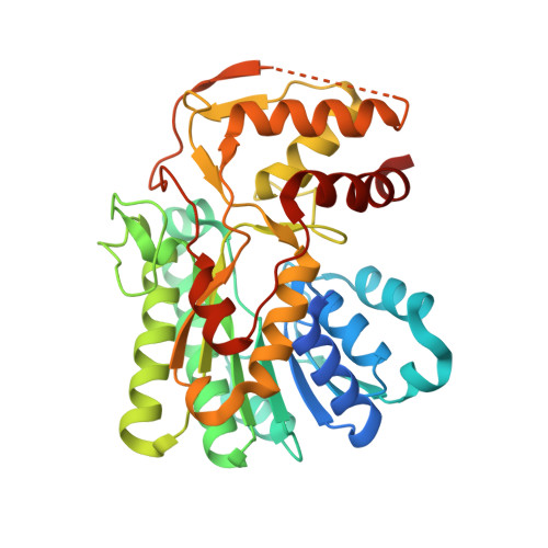
3LU1

Crystal Structure Analysis of WbgU: a UDP-GalNAc 4-epimerase


Experimental Data Snapshot

  • Method: X-RAY DIFFRACTION
  • Resolution: 2.50 Å
  • R-Value Free: 0.258 
  • R-Value Work: 0.204 
  • R-Value Observed: 0.207 

wwPDB Validation   3D Report Full Report


Ligand Structure Quality Assessment 


This is version 1.4 of the entry. See complete history


Literature

Altered architecture of substrate binding region defines the unique specificity of UDP-GalNAc 4-epimerases.

Bhatt, V.S.Guo, C.Y.Guan, W.Zhao, G.Yi, W.Liu, Z.J.Wang, P.G.

(2011) Protein Sci 20: 856-866

  • DOI: https://doi.org/10.1002/pro.611
  • Primary Citation of Related Structures:  
    3LU1

  • PubMed Abstract: 

    UDP-hexose 4-epimerases play a pivotal role in lipopolysaccharide (LPS) biosynthesis and Leloir pathway. These epimerases are classified into three groups based on whether they recognize nonacetylated UDP-hexoses (Group 1), both N-acetylated and nonacetylated UDP-hexoses (Group 2) or only N-acetylated UDP-hexoses (Group 3). Although the catalysis has been investigated extensively, yet a definitive model rationalizing the substrate specificity of all the three groups on a common platform is largely lacking. In this work, we present the crystal structure of WbgU, a novel UDP-hexose 4-epimerase that belongs to the Group 3. WbgU is involved in biosynthetic pathway of the unusual glycan 2-deoxy-L-altruronic acid that is found in the LPS of the pathogen Pleisomonas shigelloides. A model that defines its substrate specificity is proposed on the basis of the active site architecture. Representatives from all the three groups are then compared to rationalize their substrate specificity. This investigation reveals that the Group 3 active site architecture is markedly different from the "conserved scaffold" of the Group 1 and the Group 2 epimerases and highlights the interactions potentially responsible for the origin of specificity of the Group 3 epimerases toward N-acetylated hexoses. This study provides a platform for further engineering of the UDP-hexose 4-epimerases, leads to a deeper understanding of the LPS biosynthesis and carbohydrate recognition by proteins. It may also have implications in development of novel antibiotics and more economic synthesis of UDP-GalNAc and downstream products such as carbohydrate based vaccines.


  • Organizational Affiliation

    Department of Biochemistry, The Ohio State University, Columbus, Ohio 43210, USA.


Macromolecules
Find similar proteins by:  (by identity cutoff)  |  3D Structure
Entity ID: 1
MoleculeChains Sequence LengthOrganismDetailsImage
WbgU
A, B, C, D
364Plesiomonas shigelloidesMutation(s): 0 
Gene Names: wbgU
EC: 5.1.3.7
UniProt
Find proteins for Q7BJX9 (Plesiomonas shigelloides)
Explore Q7BJX9 
Go to UniProtKB:  Q7BJX9
Entity Groups  
Sequence Clusters30% Identity50% Identity70% Identity90% Identity95% Identity100% Identity
UniProt GroupQ7BJX9
Sequence Annotations
Expand
  • Reference Sequence
Small Molecules
Ligands 5 Unique
IDChains Name / Formula / InChI Key2D Diagram3D Interactions
NAD
Query on NAD

Download Ideal Coordinates CCD File 
E [auth A],
H [auth B],
K [auth C],
O [auth D]
NICOTINAMIDE-ADENINE-DINUCLEOTIDE
C21 H27 N7 O14 P2
BAWFJGJZGIEFAR-NNYOXOHSSA-N
UD2
Query on UD2

Download Ideal Coordinates CCD File 
F [auth A],
I [auth B],
L [auth C],
P [auth D]
URIDINE-DIPHOSPHATE-N-ACETYLGALACTOSAMINE
C17 H27 N3 O17 P2
LFTYTUAZOPRMMI-NESSUJCYSA-N
SO4
Query on SO4

Download Ideal Coordinates CCD File 
M [auth C],
R [auth D],
S [auth D]
SULFATE ION
O4 S
QAOWNCQODCNURD-UHFFFAOYSA-L
GLY
Query on GLY

Download Ideal Coordinates CCD File 
G [auth A],
J [auth B],
N [auth C],
T [auth D]
GLYCINE
C2 H5 N O2
DHMQDGOQFOQNFH-UHFFFAOYSA-N
NA
Query on NA

Download Ideal Coordinates CCD File 
Q [auth D]SODIUM ION
Na
FKNQFGJONOIPTF-UHFFFAOYSA-N
Experimental Data & Validation

Experimental Data

  • Method: X-RAY DIFFRACTION
  • Resolution: 2.50 Å
  • R-Value Free: 0.258 
  • R-Value Work: 0.204 
  • R-Value Observed: 0.207 
  • Space Group: P 32
Unit Cell:
Length ( Å )Angle ( ˚ )
a = 78.13α = 90
b = 78.13β = 90
c = 231.924γ = 120
Software Package:
Software NamePurpose
MOSFLMdata reduction
SCALAdata scaling
PHASERphasing
REFMACrefinement
PDB_EXTRACTdata extraction

Structure Validation

View Full Validation Report


Ligand Structure Quality Assessment 


Entry History 

Deposition Data

Revision History  (Full details and data files)

  • Version 1.0: 2010-07-21
    Type: Initial release
  • Version 1.1: 2011-07-13
    Changes: Version format compliance
  • Version 1.2: 2014-11-12
    Changes: Structure summary
  • Version 1.3: 2017-11-01
    Changes: Refinement description
  • Version 1.4: 2023-09-06
    Changes: Data collection, Database references, Derived calculations, Refinement description