3LQ2

E. coli pyruvate dehydrogenase complex E1 E235A mutant with low TDP concentration


Experimental Data Snapshot

  • Method: X-RAY DIFFRACTION
  • Resolution: 1.96 Å
  • R-Value Free: 0.223 
  • R-Value Work: 0.194 

wwPDB Validation   3D Report Full Report


Ligand Structure Quality Assessment 


This is version 2.2 of the entry. See complete history


Literature

Communication between thiamin cofactors in the Escherichia coli pyruvate dehydrogenase complex E1 component active centers: evidence for a "direct pathway" between the 4'-aminopyrimidine N1' atoms.

Nemeria, N.S.Arjunan, P.Chandrasekhar, K.Mossad, M.Tittmann, K.Furey, W.Jordan, F.

(2010) J Biol Chem 285: 11197-11209

  • DOI: https://doi.org/10.1074/jbc.M109.069179
  • Primary Citation of Related Structures:  
    3LPL, 3LQ2, 3LQ4

  • PubMed Abstract: 

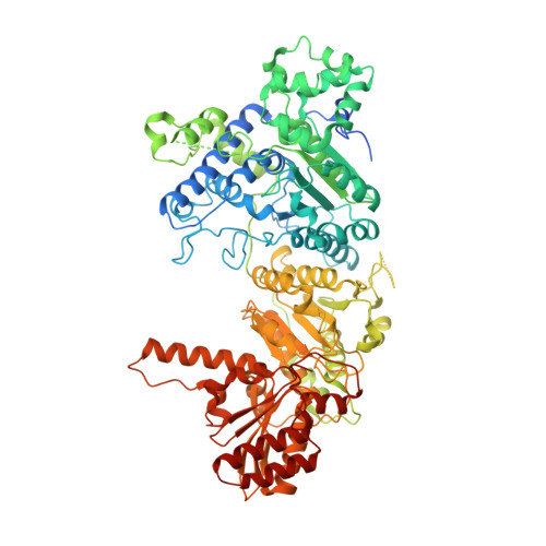
    Kinetic, spectroscopic, and structural analysis tested the hypothesis that a chain of residues connecting the 4'-aminopyrimidine N1' atoms of thiamin diphosphates (ThDPs) in the two active centers of the Escherichia coli pyruvate dehydrogenase complex E1 component provides a signal transduction pathway. Substitution of the three acidic residues (Glu(571), Glu(235), and Glu(237)) and Arg(606) resulted in impaired binding of the second ThDP, once the first active center was filled, suggesting a pathway for communication between the two ThDPs. 1) Steady-state kinetic and fluorescence quenching studies revealed that upon E571A, E235A, E237A, and R606A substitutions, ThDP binding in the second active center was affected. 2) Analysis of the kinetics of thiazolium C2 hydrogen/deuterium exchange of enzyme-bound ThDP suggests half-of-the-sites reactivity for the E1 component, with fast (activated site) and slow exchanging sites (dormant site). The E235A and E571A variants gave no evidence for the slow exchanging site, indicating that only one of two active sites is filled with ThDP. 3) Titration of the E235A and E237A variants with methyl acetylphosphonate monitored by circular dichroism suggested that only half of the active sites were filled with a covalent predecarboxylation intermediate analog. 4) Crystal structures of E235A and E571A in complex with ThDP revealed the structural basis for the spectroscopic and kinetic observations and showed that either substitution affects cofactor binding, despite the fact that Glu(235) makes no direct contact with the cofactor. The role of the conserved Glu(571) residue in both catalysis and cofactor orientation is revealed by the combined results for the first time.


  • Organizational Affiliation

    Department of Chemistry, Rutgers University, Newark, New Jersey 07102, USA.


Macromolecules
Find similar proteins by:  (by identity cutoff)  |  3D Structure
Entity ID: 1
MoleculeChains Sequence LengthOrganismDetailsImage
Pyruvate dehydrogenase E1 component
A, B
886Escherichia coli O157:H7Mutation(s): 1 
Gene Names: aceEB0114ECs0118Z0124
EC: 1.2.4.1
UniProt
Find proteins for P0AFG9 (Escherichia coli O157:H7)
Explore P0AFG9 
Go to UniProtKB:  P0AFG9
Entity Groups  
Sequence Clusters30% Identity50% Identity70% Identity90% Identity95% Identity100% Identity
UniProt GroupP0AFG9
Sequence Annotations
Expand
  • Reference Sequence
Small Molecules
Ligands 4 Unique
IDChains Name / Formula / InChI Key2D Diagram3D Interactions
TPP
Query on TPP

Download Ideal Coordinates CCD File 
C [auth A],
G [auth B]
THIAMINE DIPHOSPHATE
C12 H19 N4 O7 P2 S
AYEKOFBPNLCAJY-UHFFFAOYSA-O
EPE
Query on EPE

Download Ideal Coordinates CCD File 
F [auth A]4-(2-HYDROXYETHYL)-1-PIPERAZINE ETHANESULFONIC ACID
C8 H18 N2 O4 S
JKMHFZQWWAIEOD-UHFFFAOYSA-N
PO4
Query on PO4

Download Ideal Coordinates CCD File 
E [auth A],
I [auth B]
PHOSPHATE ION
O4 P
NBIIXXVUZAFLBC-UHFFFAOYSA-K
MG
Query on MG

Download Ideal Coordinates CCD File 
D [auth A],
H [auth B]
MAGNESIUM ION
Mg
JLVVSXFLKOJNIY-UHFFFAOYSA-N
Experimental Data & Validation

Experimental Data

  • Method: X-RAY DIFFRACTION
  • Resolution: 1.96 Å
  • R-Value Free: 0.223 
  • R-Value Work: 0.194 
  • Space Group: P 1 21 1
Unit Cell:
Length ( Å )Angle ( ˚ )
a = 80.65α = 90
b = 141.77β = 103.36
c = 83.42γ = 90
Software Package:
Software NamePurpose
CNSrefinement
PDB_EXTRACTdata extraction
HKL-2000data reduction
SCALEPACKdata scaling
PHASESphasing

Structure Validation

View Full Validation Report


Ligand Structure Quality Assessment 


Entry History 

Deposition Data

  • Released Date: 2010-03-02 
  • Deposition Author(s): Furey, W.

Revision History  (Full details and data files)

  • Version 1.0: 2010-03-02
    Type: Initial release
  • Version 1.1: 2011-07-13
    Changes: Version format compliance
  • Version 2.0: 2021-08-04
    Changes: Atomic model, Data collection, Derived calculations, Non-polymer description, Structure summary
  • Version 2.1: 2021-10-13
    Changes: Database references
  • Version 2.2: 2023-09-06
    Changes: Data collection, Refinement description