3LPV | pdb_00003lpv

X-ray crystal structure of duplex DNA containing a cisplatin 1,2-d(GpG) intrastrand cross-link

  • Classification: DNA
  • Mutation(s): No 

  • Deposited: 2010-02-06 Released: 2010-08-11 
  • Deposition Author(s): Todd, R.C., Lippard, S.J.

Experimental Data Snapshot

  • Method: X-RAY DIFFRACTION
  • Resolution: 1.77 Å
  • R-Value Free: 
    0.198 (Depositor), 0.199 (DCC) 
  • R-Value Work: 
    0.172 (Depositor), 0.175 (DCC) 
  • R-Value Observed: 
    0.173 (Depositor) 

Starting Model: experimental
View more details

wwPDB Validation 3D Report Full Report

Validation slider image for 3LPV

This is version 1.3 of the entry. See complete history

Literature

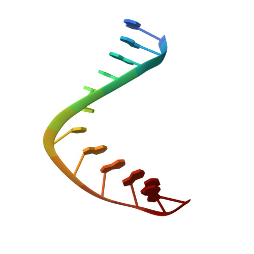
Structure of duplex DNA containing the cisplatin 1,2-{Pt(NH(3))(2)}(2+)-d(GpG) cross-link at 1.77A resolution.

Todd, R.C.Lippard, S.J.

(2010) J Inorg Biochem 104: 902-908

  • DOI: https://doi.org/10.1016/j.jinorgbio.2010.04.005
  • Primary Citation Related Structures: 
    3LPV

  • PubMed Abstract: 

    We report the 1.77-A resolution X-ray crystal structure of a dodecamer DNA duplex with the sequence 5'-CCTCTGGTCTCC-3' that has been modified to contain a single engineered 1,2-cis-{Pt(NH(3))(2)}(2+)-d(GpG) cross-link, the major DNA adduct of cisplatin. These data represent a significant improvement in resolution over the previously published 2.6-A structure. The ammine ligands in this structure are clearly resolved, leading to improved visualization of the cross-link geometry with respect to both the platinum center and to the nucleobases, which adopt a higher energy conformation. Also better resolved are the deoxyribose sugar puckers, which allow us to re-examine the global structure of platinum-modified DNA. Another new feature of this model is the location of four octahedral [Mg(H(2)O)(6)](2+) ions associated with bases in the DNA major groove and the identification of 124 ordered water molecules that participate in hydrogen-bonding interactions with either the nucleic acid or the diammineplatinum(II) moiety.


  • Organizational Affiliation
    • Department of Chemistry, Massachusetts Institute of Technology, 77 Massachusetts Avenue, Cambridge, MA 02139, United States.

Macromolecule Content 

  • Total Structure Weight: 15.35 kDa 
  • Atom Count: 1,130 
  • Modeled Residue Count: 48 
  • Deposited Residue Count: 48 
  • Unique nucleic acid chains: 2

Macromolecules

Find similar nucleic acids by:  Sequence
Entity ID: 1
MoleculeChains LengthOrganismImage
5'-D(*CP*CP*TP*CP*TP*GP*GP*TP*CP*TP*CP*C)-3'
A, C
12N/A
Sequence Annotations
Expand
Reference Sequence
Find similar nucleic acids by:  Sequence
Entity ID: 2
MoleculeChains LengthOrganismImage
5'-D(*GP*GP*AP*GP*AP*CP*CP*AP*GP*AP*GP*G)-3'
B, D
12N/A
Sequence Annotations
Expand
Reference Sequence

Experimental Data & Validation

Experimental Data

  • Method: X-RAY DIFFRACTION
  • Resolution: 1.77 Å
  • R-Value Free:  0.198 (Depositor), 0.199 (DCC) 
  • R-Value Work:  0.172 (Depositor), 0.175 (DCC) 
  • R-Value Observed: 0.173 (Depositor) 
Space Group: P 1
Unit Cell:
Length ( Å )Angle ( ˚ )
a = 31.303α = 80.06
b = 35.425β = 84.09
c = 45.128γ = 81.77
Software Package:
Software NamePurpose
DENZOdata reduction
SCALEPACKdata scaling
PHASERphasing
REFMACrefinement
PDB_EXTRACTdata extraction
HKL-2000data collection
HKL-2000data reduction
HKL-2000data scaling

Structure Validation

View Full Validation Report


Entry History 

Deposition Data

Revision History  (Full details and data files)

  • Version 1.0: 2010-08-11
    Type: Initial release
  • Version 1.1: 2011-07-13
    Changes: Version format compliance
  • Version 1.2: 2017-11-01
    Changes: Refinement description
  • Version 1.3: 2023-09-06
    Changes: Data collection, Database references, Derived calculations, Refinement description