3LM9

Crystal structure of fructokinase with ADP and Fructose bound in the active site


Experimental Data Snapshot

  • Method: X-RAY DIFFRACTION
  • Resolution: 2.45 Å
  • R-Value Free: 0.196 
  • R-Value Work: 0.157 
  • R-Value Observed: 0.159 

wwPDB Validation   3D Report Full Report


Ligand Structure Quality Assessment 


This is version 1.4 of the entry. See complete history


Literature

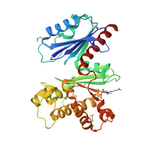
Structural studies of ROK fructokinase YdhR from Bacillus subtilis: insights into substrate binding and fructose specificity.

Nocek, B.Stein, A.J.Jedrzejczak, R.Cuff, M.E.Li, H.Volkart, L.Joachimiak, A.

(2011) J Mol Biol 406: 325-342

  • DOI: https://doi.org/10.1016/j.jmb.2010.12.021
  • Primary Citation of Related Structures:  
    1XC3, 3LM9, 3OHR

  • PubMed Abstract: 
  • The main pathway of bacterial sugar phosphorylation utilizes specific phosphoenolpyruvate phosphotransferase system (PTS) enzymes. In addition to the classic PTS system, a PTS-independent secondary system has been described in which nucleotide-dependent sugar kinases are used for monosaccharide phosphorylation ...

    The main pathway of bacterial sugar phosphorylation utilizes specific phosphoenolpyruvate phosphotransferase system (PTS) enzymes. In addition to the classic PTS system, a PTS-independent secondary system has been described in which nucleotide-dependent sugar kinases are used for monosaccharide phosphorylation. Fructokinase (FK), which phosphorylates d-fructose with ATP as a cofactor, has been shown to be a member of this secondary system. Bioinformatic analysis has shown that FK is a member of the "ROK" (bacterial Repressors, uncharacterized Open reading frames, and sugar Kinases) sequence family. In this study, we report the crystal structures of ROK FK from Bacillus subtilis (YdhR) (a) apo and in the presence of (b) ADP and (c) ADP/d-fructose. All structures show that YdhR is a homodimer with a monomer composed of two similar α/β domains forming a large cleft between domains that bind ADP and D-fructose. Enzymatic activity assays support YdhR function as an ATP-dependent fructose kinase.


    Organizational Affiliation

    Midwest Center for Structural Genomics and Structural Biology Center, Biosciences, Argonne National Laboratory, 9700 South Cass Avenue, Building 202, Argonne, IL 60439, USA.


Macromolecules
Find similar proteins by:  (by identity cutoff)  |  3D Structure
Entity ID: 1
MoleculeChainsSequence LengthOrganismDetailsImage
fructokinaseA302Bacillus subtilisMutation(s): 0 
Gene Names: gmuEydhRBSU05860
EC: 2.7.1.4
UniProt
Find proteins for O05510 (Bacillus subtilis (strain 168))
Explore O05510 
Go to UniProtKB:  O05510
Entity Groups  
Sequence Clusters30% Identity50% Identity70% Identity90% Identity95% Identity100% Identity
UniProt GroupO05510
Protein Feature View
Expand
  • Reference Sequence
Small Molecules
Ligands 4 Unique
IDChainsName / Formula / InChI Key2D Diagram3D Interactions
ADP
Query on ADP

Download Ideal Coordinates CCD File 
D [auth A]ADENOSINE-5'-DIPHOSPHATE
C10 H15 N5 O10 P2
XTWYTFMLZFPYCI-KQYNXXCUSA-N
 Ligand Interaction
FRU
Query on FRU

Download Ideal Coordinates CCD File 
B [auth A]beta-D-fructofuranose
C6 H12 O6
RFSUNEUAIZKAJO-ARQDHWQXSA-N
 Ligand Interaction
SO4
Query on SO4

Download Ideal Coordinates CCD File 
C [auth A]SULFATE ION
O4 S
QAOWNCQODCNURD-UHFFFAOYSA-L
 Ligand Interaction
ZN
Query on ZN

Download Ideal Coordinates CCD File 
E [auth A]ZINC ION
Zn
PTFCDOFLOPIGGS-UHFFFAOYSA-N
 Ligand Interaction
Modified Residues  2 Unique
IDChainsTypeFormula2D DiagramParent
MLY
Query on MLY
A L-PEPTIDE LINKINGC8 H18 N2 O2LYS
MSE
Query on MSE
A L-PEPTIDE LINKINGC5 H11 N O2 SeMET
Experimental Data & Validation

Experimental Data

  • Method: X-RAY DIFFRACTION
  • Resolution: 2.45 Å
  • R-Value Free: 0.196 
  • R-Value Work: 0.157 
  • R-Value Observed: 0.159 
  • Space Group: P 31 2 1
Unit Cell:
Length ( Å )Angle ( ˚ )
a = 112.333α = 90
b = 112.333β = 90
c = 73.958γ = 120
Software Package:
Software NamePurpose
SBC-Collectdata collection
MOLEMAN2model building
CCP4model building
REFMACrefinement
HKL-3000data reduction
HKL-3000data scaling
MOLEMAN2phasing
CCP4phasing

Structure Validation

View Full Validation Report


Ligand Structure Quality Assessment 


Entry History 

Deposition Data

Revision History  (Full details and data files)

  • Version 1.0: 2010-03-09
    Type: Initial release
  • Version 1.1: 2011-07-13
    Changes: Advisory, Version format compliance
  • Version 1.2: 2012-10-10
    Changes: Database references
  • Version 1.3: 2017-11-01
    Changes: Refinement description
  • Version 1.4: 2020-07-29
    Type: Remediation
    Reason: Carbohydrate remediation
    Changes: Data collection, Derived calculations, Structure summary