3LHB

THE 2.7 ANGSTROM CRYSTAL STRUCTURE OF DEOXYGENATED HEMOGLOBIN FROM THE SEA LAMPREY (PETROMYZON MARINUS)


Experimental Data Snapshot

  • Method: X-RAY DIFFRACTION
  • Resolution: 2.70 Å
  • R-Value Free: 0.232 
  • R-Value Work: 0.195 
  • R-Value Observed: 0.195 

wwPDB Validation   3D Report Full Report


Ligand Structure Quality Assessment 


This is version 1.3 of the entry. See complete history


Literature

The 2.7 A crystal structure of deoxygenated hemoglobin from the sea lamprey (Petromyzon marinus): structural basis for a lowered oxygen affinity and Bohr effect.

Heaslet, H.A.Royer Jr., W.E.

(1999) Structure 7: 517-526

  • DOI: https://doi.org/10.1016/s0969-2126(99)80068-9
  • Primary Citation of Related Structures:  
    3LHB

  • PubMed Abstract: 

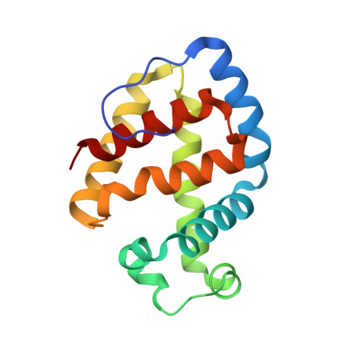
    The hemoglobins of the sea lamprey are unusual in that cooperativity and sensitivity to pH arise from an equilibrium between a high-affinity monomer and a low-affinity oligomer. Although the crystal structure of the monomeric cyanide derivative has previously been determined, the manner by which oligomerization acts to lower the oxygen affinity and confer a strong Bohr effect has, until now, been speculative. We have determined the crystal structure of deoxygenated lamprey hemoglobin V by molecular replacement to 2.7 A resolution, in a crystal form with twelve protomers in the asymmetric unit. The subunits are arranged as six essentially identical dimers, with a novel subunit interface formed by the E helices and the AB corner using the standard hemoglobin helical designations. In addition to nonpolar interactions, the interface includes a striking cluster of four glutamate residues. The proximity of the interface to ligand-binding sites implicates a direct effect on ligand affinity. Comparison of the deoxy structure with that of the cyanide derivative revealed conformational changes that appear to be linked to the functional behavior. Oligomerization is coupled with a movement of the first half of the E helix by up to 1.0 A towards the heme, resulting in steric interference of ligand binding to the deoxy structure. The Bohr effect seems to result from proton uptake by glutamate residues as they are buried in the interface. Unlike human and mollusc hemoglobins, in which modulation of function is due to primarily proximal effects, regulation of oxygen affinity in lamprey hemoglobin V seems to depend on changes at the distal (ligand-binding) side of the heme group.


  • Organizational Affiliation

    Department of Biochemistry and Molecular Biology, University of Massachusetts Medical School, Worcester 01655, USA.


Macromolecules
Find similar proteins by:  (by identity cutoff)  |  3D Structure
Entity ID: 1
MoleculeChains Sequence LengthOrganismDetailsImage
PROTEIN (HEMOGLOBIN)
A, B, C, D, E
A, B, C, D, E, F, G, H, I, J, K, L
149Petromyzon marinusMutation(s): 0 
UniProt
Find proteins for P02208 (Petromyzon marinus)
Explore P02208 
Go to UniProtKB:  P02208
Entity Groups  
Sequence Clusters30% Identity50% Identity70% Identity90% Identity95% Identity100% Identity
UniProt GroupP02208
Sequence Annotations
Expand
  • Reference Sequence
Small Molecules
Ligands 1 Unique
IDChains Name / Formula / InChI Key2D Diagram3D Interactions
HEM
Query on HEM

Download Ideal Coordinates CCD File 
M [auth A]
N [auth B]
O [auth C]
P [auth D]
Q [auth E]
M [auth A],
N [auth B],
O [auth C],
P [auth D],
Q [auth E],
R [auth F],
S [auth G],
T [auth H],
U [auth I],
V [auth J],
W [auth K],
X [auth L]
PROTOPORPHYRIN IX CONTAINING FE
C34 H32 Fe N4 O4
KABFMIBPWCXCRK-RGGAHWMASA-L
Experimental Data & Validation

Experimental Data

  • Method: X-RAY DIFFRACTION
  • Resolution: 2.70 Å
  • R-Value Free: 0.232 
  • R-Value Work: 0.195 
  • R-Value Observed: 0.195 
  • Space Group: P 1 21 1
Unit Cell:
Length ( Å )Angle ( ˚ )
a = 59.63α = 90
b = 215.16β = 95.8
c = 75.39γ = 90
Software Package:
Software NamePurpose
DENZOdata reduction
SCALEPACKdata scaling
AMoREphasing
X-PLORrefinement

Structure Validation

View Full Validation Report


Ligand Structure Quality Assessment 


Entry History 

Deposition Data

Revision History  (Full details and data files)

  • Version 1.0: 1999-01-20
    Type: Initial release
  • Version 1.1: 2008-04-27
    Changes: Version format compliance
  • Version 1.2: 2011-07-13
    Changes: Version format compliance
  • Version 1.3: 2023-09-06
    Changes: Data collection, Database references, Derived calculations, Refinement description