


Crystal structure of SusD-like carbohydrate binding protein (YP_001298396.1) from Bacteroides vulgatus ATCC 8482 at 1.70 A resolution
Joint Center for Structural Genomics (JCSG)To be published.
Experimental Data Snapshot
wwPDB Validation 3D Report Full Report

Entity ID: 1 | |||||
|---|---|---|---|---|---|
| Molecule | Chains | Sequence Length | Organism | Details | Image |
| SusD-like carbohydrate binding protein | 495 | Phocaeicola vulgatus ATCC 8482 | Mutation(s): 0 Gene Names: BVU_1083 | 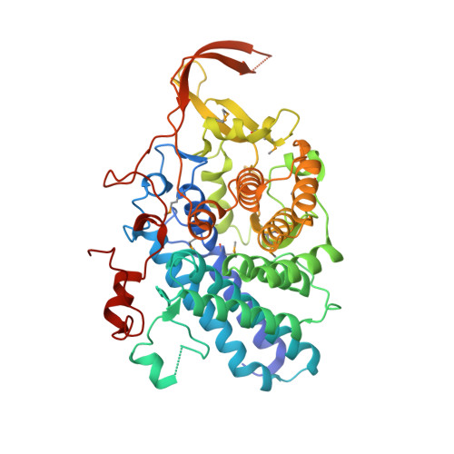 | |
UniProt | |||||
Entity Groups | |||||
| Sequence Clusters | 30% Identity50% Identity70% Identity90% Identity95% Identity100% Identity | ||||
| UniProt Group | A6KZB0 | ||||
Sequence AnnotationsExpand | |||||
Reference Sequence | |||||
| Ligands 2 Unique | |||||
|---|---|---|---|---|---|
| ID | Chains | Name / Formula / InChI Key | 2D Diagram | 3D Interactions | |
| 2PE Download:Ideal Coordinates CCD File | E [auth A] | NONAETHYLENE GLYCOL C18 H38 O10 YZUUTMGDONTGTN-UHFFFAOYSA-N |  | ||
| 1PE Download:Ideal Coordinates CCD File | B [auth A] C [auth A] D [auth A] F [auth A] G [auth A] | PENTAETHYLENE GLYCOL C10 H22 O6 JLFNLZLINWHATN-UHFFFAOYSA-N |  | ||
| Modified Residues 1 Unique | |||||
|---|---|---|---|---|---|
| ID | Chains | Type | Formula | 2D Diagram | Parent |
| MSE Query on MSE | A | L-PEPTIDE LINKING | C5 H11 N O2 Se |  | MET |
| Length ( Å ) | Angle ( ˚ ) |
|---|---|
| a = 191.772 | α = 90 |
| b = 46.707 | β = 106.55 |
| c = 73.524 | γ = 90 |
| Software Name | Purpose |
|---|---|
| REFMAC | refinement |
| PHENIX | refinement |
| SHELX | phasing |
| MolProbity | model building |
| XSCALE | data scaling |
| PDB_EXTRACT | data extraction |
| XDS | data reduction |
| SHELXD | phasing |
| autoSHARP | phasing |


