3L8L

Gramicidin D complex with sodium iodide


Experimental Data Snapshot

  • Method: X-RAY DIFFRACTION
  • Resolution: 1.25 Å

wwPDB Validation   3D Report Full Report


This is version 2.0 of the entry. See complete history


Literature

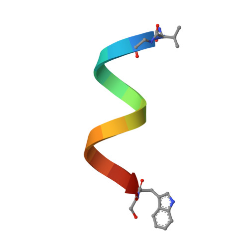
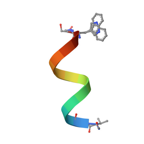
The first crystal structure of a gramicidin complex with sodium: high-resolution study of a nonstoichiometric gramicidin D-NaI complex.

Olczak, A.Glowka, M.L.Szczesio, M.Bojarska, J.Wawrzak, Z.Duax, W.L.

(2010) Acta Crystallogr D Biol Crystallogr 66: 874-880

  • DOI: https://doi.org/10.1107/S0907444910019876
  • Primary Citation of Related Structures:  
    3L8L

  • PubMed Abstract: 

    The crystal structure of the nonstoichiometric complex of gramicidin D with NaI has been studied using synchrotron radiation at 100 K. The limiting resolution was 1.25 A and the R factor was 16% for 19 883 observed reflections. The general architecture of the antiparallel two-stranded gramicidin dimers in the studied crystal was a right-handed antiparallel double-stranded form that closely resembles the structures of other right-handed species published to date. However, there were several surprising observations. In addition to the significantly different composition of linear gramicidins identified in the crystal structure, including the absence of the gramicidin C form, only two cationic sites were found in each of the two independent dimers (channels), which were partially occupied by sodium, compared with the seven sites found in the RbCl complex of gramicidin. The sum of the partial occupancies of Na(+) was only 1.26 per two dimers and was confirmed by the similar content of iodine ions (1.21 ions distributed over seven sites), which was easily visible from their anomalous signal. Another surprising observation was the significant asymmetry of the distributions and occupancies of cations in the gramicidin dimers, which was in contrast to those observed in the high-resolution structures of the complexes of heavier alkali metals with gramicidin D, especially that of rubidium.


  • Organizational Affiliation

    Institute of General and Ecological Chemistry, Technical University of Łódź, Zeromskiego 116, 90-924 Łódź, Poland. olczakan@p.lodz.pl


Macromolecules

Find similar proteins by:  Sequence   |   3D Structure  

Entity ID: 1
MoleculeChains Sequence LengthOrganismDetailsImage
GRAMICIDIN D
A, C
16Brevibacillus brevisMutation(s): 0 
Entity Groups  
Sequence Clusters30% Identity50% Identity70% Identity90% Identity95% Identity100% Identity
Sequence Annotations
Expand
  • Reference Sequence

Find similar proteins by:  Sequence   |   3D Structure  

Entity ID: 2
MoleculeChains Sequence LengthOrganismDetailsImage
GRAMICIDIN D
B, D
16Brevibacillus brevisMutation(s): 1 
Entity Groups  
Sequence Clusters30% Identity50% Identity70% Identity90% Identity95% Identity100% Identity
Sequence Annotations
Expand
  • Reference Sequence
Small Molecules
Ligands 3 Unique
IDChains Name / Formula / InChI Key2D Diagram3D Interactions
IOD
Query on IOD

Download Ideal Coordinates CCD File 
F [auth A]
G [auth A]
H [auth A]
K [auth B]
O [auth C]
F [auth A],
G [auth A],
H [auth A],
K [auth B],
O [auth C],
Q [auth D],
R [auth D]
IODIDE ION
I
XMBWDFGMSWQBCA-UHFFFAOYSA-M
MOH
Query on MOH

Download Ideal Coordinates CCD File 
I [auth A],
L [auth B],
M [auth B]
METHANOL
C H4 O
OKKJLVBELUTLKV-UHFFFAOYSA-N
NA
Query on NA

Download Ideal Coordinates CCD File 
E [auth A],
J [auth B],
N [auth C],
P [auth D]
SODIUM ION
Na
FKNQFGJONOIPTF-UHFFFAOYSA-N
Modified Residues  1 Unique
IDChains TypeFormula2D DiagramParent
FVA
Query on FVA
A, C
L-PEPTIDE LINKINGC6 H11 N O3VAL
Experimental Data & Validation

Experimental Data

  • Method: X-RAY DIFFRACTION
  • Resolution: 1.25 Å
  • Space Group: P 21 21 21
Unit Cell:
Length ( Å )Angle ( ˚ )
a = 29.816α = 90
b = 31.255β = 90
c = 51.667γ = 90
Software Package:
Software NamePurpose
AMoREphasing
SHELXL-97refinement
DENZOdata reduction
SCALEPACKdata scaling

Structure Validation

View Full Validation Report


Entry History 

Deposition Data

Revision History  (Full details and data files)

  • Version 1.0: 2010-07-28
    Type: Initial release
  • Version 1.1: 2011-06-14
    Changes: Version format compliance
  • Version 1.2: 2011-07-13
    Changes: Version format compliance
  • Version 1.3: 2011-07-27
    Changes: Atomic model, Database references, Derived calculations, Non-polymer description, Structure summary
  • Version 1.4: 2012-03-28
    Changes: Database references
  • Version 1.5: 2012-12-12
    Changes: Other
  • Version 1.6: 2013-02-13
    Changes: Derived calculations
  • Version 1.7: 2023-09-06
    Changes: Data collection, Database references, Derived calculations, Refinement description
  • Version 2.0: 2023-11-15
    Changes: Atomic model, Data collection, Derived calculations