Structural Determinants of Substrate Recognition in the HAD Superfamily Member d-glycero-d-manno-Heptose-1,7-bisphosphate Phosphatase (GmhB) .
Nguyen, H.H., Wang, L., Huang, H., Peisach, E., Dunaway-Mariano, D., Allen, K.N.(2010) Biochemistry 49: 1082-1092
- PubMed: 20050614 Search on PubMedSearch on PubMed Central
- DOI: https://doi.org/10.1021/bi902019q
- Primary Citation Related Structures:
3L8E, 3L8F, 3L8G, 3L8H - PubMed Abstract:
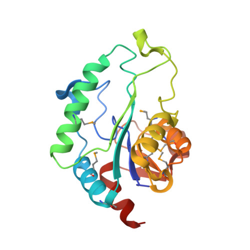
The haloalkanoic acid dehalogenase (HAD) enzyme superfamily is the largest family of phosphohydrolases. In HAD members, the structural elements that provide the binding interactions that support substrate specificity are separated from those that orchestrate catalysis. For most HAD phosphatases, a cap domain functions in substrate recognition. However, for the HAD phosphatases that lack a cap domain, an alternate strategy for substrate selection must be operative. One such HAD phosphatase, GmhB of the HisB subfamily, was selected for structure-function analysis. Herein, the X-ray crystallographic structures of Escherichia coli GmhB in the apo form (1.6 A resolution), in a complex with Mg(2+) and orthophosphate (1.8 A resolution), and in a complex with Mg(2+) and d-glycero-d-manno-heptose 1beta,7-bisphosphate (2.2 A resolution) were determined, in addition to the structure of Bordetella bronchiseptica GmhB bound to Mg(2+) and orthophosphate (1.7 A resolution). The structures show that in place of a cap domain, the GmhB catalytic site is elaborated by three peptide inserts or loops that pack to form a concave, semicircular surface around the substrate leaving group. Structure-guided kinetic analysis of site-directed mutants was conducted in parallel with a bioinformatics study of sequence diversification within the HisB subfamily to identify loop residues that serve as substrate recognition elements and that distinguish GmhB from its subfamily counterpart, the histidinol-phosphate phosphatase domain of HisB. We show that GmhB and the histidinol-phosphate phosphatase domain use the same design of three substrate recognition loops inserted into the cap domain yet, through selective residue usage on the loops, have achieved unique substrate specificity and thus novel biochemical function.
- Department of Physiology and Biophysics, Boston University School of Medicine, Boston, Massachusetts 02118-2394, USA.
Organizational Affiliation: