3L32 | pdb_00003l32

Structure of the dimerisation domain of the rabies virus phosphoprotein


Experimental Data Snapshot

  • Method: X-RAY DIFFRACTION
  • Resolution: 1.50 Å
  • R-Value Free: 
    0.228 (Depositor), 0.232 (DCC) 
  • R-Value Work: 
    0.196 (Depositor), 0.197 (DCC) 
  • R-Value Observed: 
    0.198 (Depositor) 

wwPDB Validation 3D Report Full Report

Validation slider image for 3L32

This is version 1.2 of the entry. See complete history

Literature

Structure of the dimerisation domain of the rabies virus phosphoprotein

Ivanov, I.Crepin, T.Jamin, M.Ruigrok, R.W.H.

(2010) J Virol 

  • DOI: https://doi.org/10.1128/JVI.02557-09
  • Primary Citation Related Structures: 
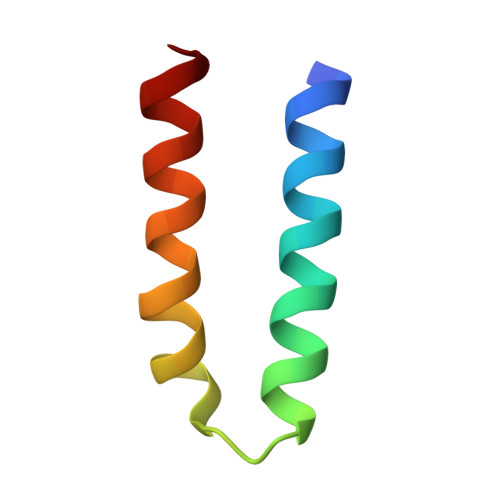
    3L32

  • PubMed Abstract: 

    The crystal structure of the dimerization domain of rabies virus phosphoprotein was determined. The monomer consists of two alpha-helices that make a helical hairpin held together mainly by hydrophobic interactions. The monomer has a hydrophilic and a hydrophobic face, and in the dimer two monomers pack together through their hydrophobic surfaces. This structure is very different from the dimerization domain of the vesicular stomatitis virus phosphoprotein and also from the tetramerization domain of the Sendai virus phosphoprotein, suggesting that oligomerization is conserved but not structure.


  • Organizational Affiliation
    • UVHCI, UMI 3265 UJF-EMBL-CNRS, BP 181, 38042 Grenoble, Cedex 9, France.

Macromolecule Content 

  • Total Structure Weight: 10.69 kDa 
  • Atom Count: 811 
  • Modeled Residue Count: 89 
  • Deposited Residue Count: 90 
  • Unique protein chains: 1

Macromolecules

Find similar proteins by:|  3D Structure
Entity ID: 1
MoleculeChains  Sequence LengthOrganismDetailsImage
Phosphoprotein
A, B
45Rabies virus China/MRVMutation(s): 0 
Gene Names: P
UniProt
Find proteins for Q0GBY3 (Rabies virus (strain China/MRV))
Explore Q0GBY3 
Go to UniProtKB:  Q0GBY3
Entity Groups
Sequence Clusters30% Identity50% Identity70% Identity90% Identity95% Identity100% Identity
UniProt GroupQ0GBY3
Sequence Annotations
Expand
Reference Sequence

Experimental Data & Validation

Experimental Data

  • Method: X-RAY DIFFRACTION
  • Resolution: 1.50 Å
  • R-Value Free:  0.228 (Depositor), 0.232 (DCC) 
  • R-Value Work:  0.196 (Depositor), 0.197 (DCC) 
  • R-Value Observed: 0.198 (Depositor) 
Space Group: I 41 2 2
Unit Cell:
Length ( Å )Angle ( ˚ )
a = 43.29α = 90
b = 43.29β = 90
c = 192.02γ = 90
Software Package:
Software NamePurpose
REFMACrefinement
PDB_EXTRACTdata extraction
XSCALEdata scaling

Structure Validation

View Full Validation Report


Entry History 

Deposition Data

Revision History  (Full details and data files)

  • Version 1.0: 2010-02-16
    Type: Initial release
  • Version 1.1: 2011-07-13
    Changes: Version format compliance
  • Version 1.2: 2024-03-20
    Changes: Data collection, Database references