3L1M

Crystal Structure of a Ni-directed Dimer of Cytochrome cb562 with a Quinolate-Histidine Hybrid Coordination Motif


Experimental Data Snapshot

  • Method: X-RAY DIFFRACTION
  • Resolution: 2.30 Å
  • R-Value Free: 0.314 
  • R-Value Work: 0.266 
  • R-Value Observed: 0.270 

wwPDB Validation   3D Report Full Report


Ligand Structure Quality Assessment 


This is version 1.4 of the entry. See complete history


Literature

Controlled protein dimerization through hybrid coordination motifs.

Radford, R.J.Nguyen, P.C.Ditri, T.B.Figueroa, J.S.Tezcan, F.A.

(2010) Inorg Chem 49: 4362-4369

  • DOI: https://doi.org/10.1021/ic100534y
  • Primary Citation of Related Structures:  
    3L1M

  • PubMed Abstract: 

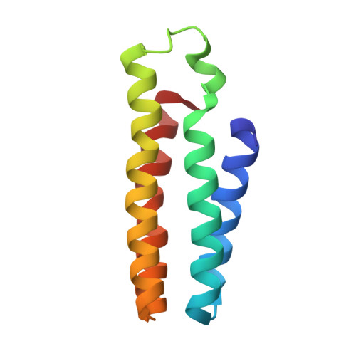
    Protein homodimerization is the simplest form of oligomerization that is frequently utilized for the construction of functional biological assemblies and the regulation of cellular pathways. Despite its simplicity, dimerization still poses an enormous challenge for protein engineering and chemical manipulation, owing to the large molecular surfaces involved in this process. We report here the construction of a hybrid coordination motif--consisting of a natural (His) and a non-natural ligand (quinolate)--on the alpha-helical surface of cytochrome cb(562), which (a) simultaneously binds divalent metals with high affinity, (b) leads to a metal-induced increase in global protein stability, and importantly, (c) enables the formation of a discrete protein dimer, whose shape is dictated by the inner-sphere metal coordination geometry and closely approximates that of the DNA-binding domains of bZIP family transcription factors.


  • Organizational Affiliation

    Department of Chemistry and Biochemistry, University of California, San Diego, 9500 Gilman Ave, La Jolla, California 92093-0356, USA.


Macromolecules
Find similar proteins by:  (by identity cutoff)  |  3D Structure
Entity ID: 1
MoleculeChains Sequence LengthOrganismDetailsImage
Soluble cytochrome b562106Escherichia coliMutation(s): 4 
UniProt
Find proteins for P0ABE7 (Escherichia coli)
Explore P0ABE7 
Go to UniProtKB:  P0ABE7
Entity Groups  
Sequence Clusters30% Identity50% Identity70% Identity90% Identity95% Identity100% Identity
UniProt GroupP0ABE7
Sequence Annotations
Expand
  • Reference Sequence
Small Molecules
Ligands 3 Unique
IDChains Name / Formula / InChI Key2D Diagram3D Interactions
HEM
Query on HEM

Download Ideal Coordinates CCD File 
B [auth A]PROTOPORPHYRIN IX CONTAINING FE
C34 H32 Fe N4 O4
KABFMIBPWCXCRK-RGGAHWMASA-L
HQI
Query on HQI

Download Ideal Coordinates CCD File 
C [auth A]N-(8-hydroxyquinolin-5-yl)acetamide
C11 H10 N2 O2
YATHWKGKLIXEFF-UHFFFAOYSA-N
NI
Query on NI

Download Ideal Coordinates CCD File 
D [auth A]NICKEL (II) ION
Ni
VEQPNABPJHWNSG-UHFFFAOYSA-N
Experimental Data & Validation

Experimental Data

  • Method: X-RAY DIFFRACTION
  • Resolution: 2.30 Å
  • R-Value Free: 0.314 
  • R-Value Work: 0.266 
  • R-Value Observed: 0.270 
  • Space Group: P 21 21 2
Unit Cell:
Length ( Å )Angle ( ˚ )
a = 88.44α = 90
b = 34.511β = 90
c = 46.896γ = 90
Software Package:
Software NamePurpose
CNSrefinement
PDB_EXTRACTdata extraction
APEXdata collection
SAINTdata reduction
SADABSdata scaling
MOLREPphasing

Structure Validation

View Full Validation Report


Ligand Structure Quality Assessment 


Entry History 

Deposition Data

Revision History  (Full details and data files)

  • Version 1.0: 2010-04-28
    Type: Initial release
  • Version 1.1: 2011-07-13
    Changes: Version format compliance
  • Version 1.2: 2018-08-29
    Changes: Advisory, Data collection, Source and taxonomy, Structure summary
  • Version 1.3: 2021-10-13
    Changes: Advisory, Database references, Derived calculations
  • Version 1.4: 2023-09-06
    Changes: Data collection, Refinement description