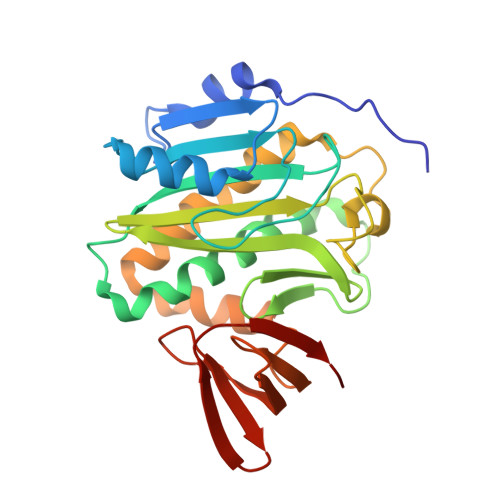
Structural ordering of disordered ligand-binding loops of biotin protein ligase into active conformations as a consequence of dehydration.
Gupta, V., Gupta, R.K., Khare, G., Salunke, D.M., Surolia, A., Tyagi, A.K.(2010) PLoS One 5: e9222-e9222
- PubMed: 20169168 Search on PubMedSearch on PubMed Central
- DOI: https://doi.org/10.1371/journal.pone.0009222
- Primary Citation Related Structures:
3L1A, 3L2Z - PubMed Abstract:
Mycobacterium tuberculosis (Mtb), a dreaded pathogen, has a unique cell envelope composed of high fatty acid content that plays a crucial role in its pathogenesis. Acetyl Coenzyme A Carboxylase (ACC), an important enzyme that catalyzes the first reaction of fatty acid biosynthesis, is biotinylated by biotin acetyl-CoA carboxylase ligase (BirA). The ligand-binding loops in all known apo BirAs to date are disordered and attain an ordered structure only after undergoing a conformational change upon ligand-binding. Here, we report that dehydration of Mtb-BirA crystals traps both the apo and active conformations in its asymmetric unit, and for the first time provides structural evidence of such transformation. Recombinant Mtb-BirA was crystallized at room temperature, and diffraction data was collected at 295 K as well as at 120 K. Transfer of crystals to paraffin and paratone-N oil (cryoprotectants) prior to flash-freezing induced lattice shrinkage and enhancement in the resolution of the X-ray diffraction data. Intriguingly, the crystal lattice rearrangement due to shrinkage in the dehydrated Mtb-BirA crystals ensued structural order of otherwise flexible ligand-binding loops L4 and L8 in apo BirA. In addition, crystal dehydration resulted in a shift of approximately 3.5 A in the flexible loop L6, a proline-rich loop unique to Mtb complex as well as around the L11 region. The shift in loop L11 in the C-terminal domain on dehydration emulates the action responsible for the complex formation with its protein ligand biotin carboxyl carrier protein (BCCP) domain of ACCA3. This is contrary to the involvement of loop L14 observed in Pyrococcus horikoshii BirA-BCCP complex. Another interesting feature that emerges from this dehydrated structure is that the two subunits A and B, though related by a noncrystallographic twofold symmetry, assemble into an asymmetric dimer representing the ligand-bound and ligand-free states of the protein, respectively. In-depth analyses of the sequence and the structure also provide answers to the reported lower affinities of Mtb-BirA toward ATP and biotin substrates. This dehydrated crystal structure not only provides key leads to the understanding of the structure/function relationships in the protein in the absence of any ligand-bound structure, but also demonstrates the merit of dehydration of crystals as an inimitable technique to have a glance at proteins in action.
- Department of Biochemistry, University of Delhi, New Delhi, India.
Organizational Affiliation: