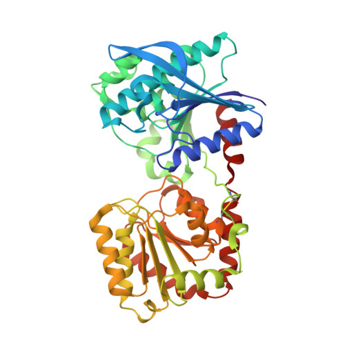
3L01

Crystal structure of monomeric glycogen synthase from Pyrococcus abyssi


Experimental Data Snapshot

  • Method: X-RAY DIFFRACTION
  • Resolution: 2.60 Å
  • R-Value Free: 0.228 
  • R-Value Work: 0.198 
  • R-Value Observed: 0.199 

Starting Model: experimental
View more details

wwPDB Validation   3D Report Full Report


This is version 2.2 of the entry. See complete history


Literature

Processivity and Subcellular Localization of Glycogen Synthase Depend on a Non-catalytic High Affinity Glycogen-binding Site.

Diaz, A.Martinez-Pons, C.Fita, I.Ferrer, J.C.Guinovart, J.J.

(2011) J Biol Chem 286: 18505-18514

  • DOI: https://doi.org/10.1074/jbc.M111.236109
  • Primary Citation of Related Structures:  
    3L01

  • PubMed Abstract: 

    Glycogen synthase, a central enzyme in glucose metabolism, catalyzes the successive addition of α-1,4-linked glucose residues to the non-reducing end of a growing glycogen molecule. A non-catalytic glycogen-binding site, identified by x-ray crystallography on the surface of the glycogen synthase from the archaeon Pyrococcus abyssi, has been found to be functionally conserved in the eukaryotic enzymes. The disruption of this binding site in both the archaeal and the human muscle glycogen synthases has a large impact when glycogen is the acceptor substrate. Instead, the catalytic efficiency remains essentially unchanged when small oligosaccharides are used as substrates. Mutants of the human muscle enzyme with reduced affinity for glycogen also show an altered intracellular distribution and a marked decrease in their capacity to drive glycogen accumulation in vivo. The presence of a high affinity glycogen-binding site away from the active center explains not only the long-recognized strong binding of glycogen synthase to glycogen but also the processivity and the intracellular localization of the enzyme. These observations demonstrate that the glycogen-binding site is a critical regulatory element responsible for the in vivo catalytic efficiency of GS.


  • Organizational Affiliation

    Institute for Research in Biomedicine, Universitat de Barcelona, Barcelona, Spain.


Macromolecules
Find similar proteins by:  (by identity cutoff)  |  3D Structure
Entity ID: 1
MoleculeChains Sequence LengthOrganismDetailsImage
GlgA glycogen synthase
A, B
428Pyrococcus abyssiMutation(s): 1 
Gene Names: PYRAB00770PAB2292
EC: 2.4.1.21
UniProt
Find proteins for Q9V2J8 (Pyrococcus abyssi (strain GE5 / Orsay))
Explore Q9V2J8 
Go to UniProtKB:  Q9V2J8
Entity Groups  
Sequence Clusters30% Identity50% Identity70% Identity90% Identity95% Identity100% Identity
UniProt GroupQ9V2J8
Sequence Annotations
Expand
  • Reference Sequence
Oligosaccharides

Help

Entity ID: 2
MoleculeChains Length2D Diagram Glycosylation3D Interactions
alpha-D-glucopyranose-(1-4)-alpha-D-glucopyranose-(1-4)-alpha-D-glucopyranose-(1-4)-alpha-D-glucopyranose
C, D
4N/A
Glycosylation Resources
GlyTouCan:  G87171PZ
GlyCosmos:  G87171PZ
GlyGen:  G87171PZ
Small Molecules
Ligands 3 Unique
IDChains Name / Formula / InChI Key2D Diagram3D Interactions
GOL
Query on GOL

Download Ideal Coordinates CCD File 
K [auth A],
L [auth A],
S [auth B],
T [auth B]
GLYCEROL
C3 H8 O3
PEDCQBHIVMGVHV-UHFFFAOYSA-N
K
Query on K

Download Ideal Coordinates CCD File 
E [auth A]
F [auth A]
G [auth A]
M [auth B]
N [auth B]
E [auth A],
F [auth A],
G [auth A],
M [auth B],
N [auth B],
O [auth B]
POTASSIUM ION
K
NPYPAHLBTDXSSS-UHFFFAOYSA-N
CL
Query on CL

Download Ideal Coordinates CCD File 
H [auth A]
I [auth A]
J [auth A]
P [auth B]
Q [auth B]
H [auth A],
I [auth A],
J [auth A],
P [auth B],
Q [auth B],
R [auth B]
CHLORIDE ION
Cl
VEXZGXHMUGYJMC-UHFFFAOYSA-M
Biologically Interesting Molecules (External Reference) 1 Unique
Experimental Data & Validation

Experimental Data

  • Method: X-RAY DIFFRACTION
  • Resolution: 2.60 Å
  • R-Value Free: 0.228 
  • R-Value Work: 0.198 
  • R-Value Observed: 0.199 
  • Space Group: P 21 21 21
Unit Cell:
Length ( Å )Angle ( ˚ )
a = 103.604α = 90
b = 119.017β = 90
c = 141.101γ = 90
Software Package:
Software NamePurpose
REFMACrefinement
PDB_EXTRACTdata extraction
MOSFLMdata reduction
SCALAdata scaling
MOLREPphasing

Structure Validation

View Full Validation Report


Entry History 

Deposition Data

Revision History  (Full details and data files)

  • Version 1.0: 2010-12-29
    Type: Initial release
  • Version 1.1: 2011-07-13
    Changes: Version format compliance
  • Version 2.0: 2020-07-29
    Type: Remediation
    Reason: Carbohydrate remediation
    Changes: Atomic model, Data collection, Database references, Derived calculations, Non-polymer description, Structure summary
  • Version 2.1: 2021-11-10
    Changes: Database references, Structure summary
  • Version 2.2: 2023-11-01
    Changes: Data collection, Refinement description