3KV4

Structure of PHF8 in complex with histone H3


Experimental Data Snapshot

  • Method: X-RAY DIFFRACTION
  • Resolution: 2.19 Å
  • R-Value Free: 0.255 
  • R-Value Work: 0.217 
  • R-Value Observed: 0.217 

wwPDB Validation   3D Report Full Report


This is version 1.2 of the entry. See complete history


Literature

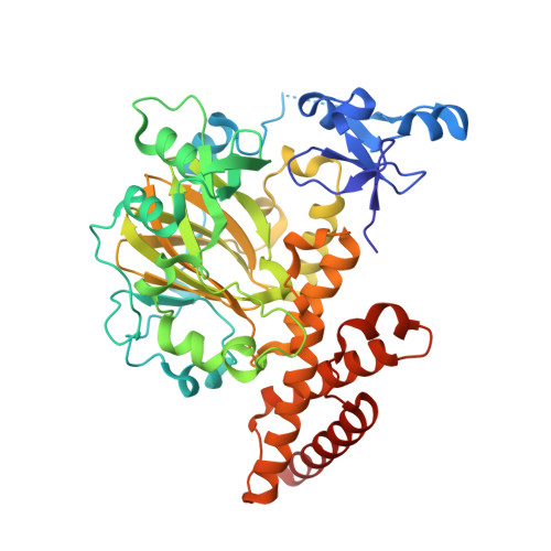
Enzymatic and structural insights for substrate specificity of a family of jumonji histone lysine demethylases.

Horton, J.R.Upadhyay, A.K.Qi, H.H.Zhang, X.Shi, Y.Cheng, X.

(2010) Nat Struct Mol Biol 17: 38-43

  • DOI: https://doi.org/10.1038/nsmb.1753
  • Primary Citation of Related Structures:  
    3KV4, 3KV5, 3KV6, 3KV9, 3KVA, 3KVB

  • PubMed Abstract: 

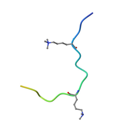
    Combinatorial readout of multiple covalent histone modifications is poorly understood. We provide insights into how an activating histone mark, in combination with linked repressive marks, is differentially 'read' by two related human demethylases, PHF8 and KIAA1718 (also known as JHDM1D). Both enzymes harbor a plant homeodomain (PHD) that binds Lys4-trimethylated histone 3 (H3K4me3) and a jumonji domain that demethylates either H3K9me2 or H3K27me2. The presence of H3K4me3 on the same peptide as H3K9me2 makes the doubly methylated peptide a markedly better substrate of PHF8, whereas the presence of H3K4me3 has the opposite effect, diminishing the H3K9me2 demethylase activity of KIAA1718 without adversely affecting its H3K27me2 activity. The difference in substrate specificity between the two is explained by PHF8 adopting a bent conformation, allowing each of its domains to engage its respective target, whereas KIAA1718 adopts an extended conformation, which prevents its access to H3K9me2 by its jumonji domain when its PHD engages H3K4me3.


  • Organizational Affiliation

    Department of Biochemistry, Emory University School of Medicine, Atlanta, Georgia, USA.


Macromolecules
Find similar proteins by:  (by identity cutoff)  |  3D Structure
Entity ID: 1
MoleculeChains Sequence LengthOrganismDetailsImage
PHD finger protein 8447Homo sapiensMutation(s): 0 
Gene Names: KIAA1111PHF8ZNF422
EC: 2
UniProt & NIH Common Fund Data Resources
Find proteins for Q9UPP1 (Homo sapiens)
Explore Q9UPP1 
Go to UniProtKB:  Q9UPP1
PHAROS:  Q9UPP1
GTEx:  ENSG00000172943 
Entity Groups  
Sequence Clusters30% Identity50% Identity70% Identity90% Identity95% Identity100% Identity
UniProt GroupQ9UPP1
Sequence Annotations
Expand
  • Reference Sequence

Find similar proteins by:  Sequence   |   3D Structure  

Entity ID: 2
MoleculeChains Sequence LengthOrganismDetailsImage
Histone H3-like24N/AMutation(s): 0 
UniProt & NIH Common Fund Data Resources
Find proteins for Q6NXT2 (Homo sapiens)
Explore Q6NXT2 
Go to UniProtKB:  Q6NXT2
PHAROS:  Q6NXT2
GTEx:  ENSG00000188375 
Entity Groups  
Sequence Clusters30% Identity50% Identity70% Identity90% Identity95% Identity100% Identity
UniProt GroupQ6NXT2
Sequence Annotations
Expand
  • Reference Sequence
Small Molecules
Ligands 5 Unique
IDChains Name / Formula / InChI Key2D Diagram3D Interactions
OGA
Query on OGA

Download Ideal Coordinates CCD File 
T [auth A]N-OXALYLGLYCINE
C4 H5 N O5
BIMZLRFONYSTPT-UHFFFAOYSA-N
ZN
Query on ZN

Download Ideal Coordinates CCD File 
C [auth A],
D [auth A]
ZINC ION
Zn
PTFCDOFLOPIGGS-UHFFFAOYSA-N
EDO
Query on EDO

Download Ideal Coordinates CCD File 
G [auth A]
H [auth A]
I [auth A]
J [auth A]
K [auth A]
G [auth A],
H [auth A],
I [auth A],
J [auth A],
K [auth A],
L [auth A],
M [auth A],
N [auth A],
O [auth A],
P [auth A],
Q [auth A],
R [auth A],
S [auth A]
1,2-ETHANEDIOL
C2 H6 O2
LYCAIKOWRPUZTN-UHFFFAOYSA-N
NI
Query on NI

Download Ideal Coordinates CCD File 
F [auth A]NICKEL (II) ION
Ni
VEQPNABPJHWNSG-UHFFFAOYSA-N
FE2
Query on FE2

Download Ideal Coordinates CCD File 
E [auth A]FE (II) ION
Fe
CWYNVVGOOAEACU-UHFFFAOYSA-N
Modified Residues  2 Unique
IDChains TypeFormula2D DiagramParent
M3L
Query on M3L
B
L-PEPTIDE LINKINGC9 H21 N2 O2LYS
MLY
Query on MLY
B
L-PEPTIDE LINKINGC8 H18 N2 O2LYS
Binding Affinity Annotations 
IDSourceBinding Affinity
OGA BindingDB:  3KV4 IC50: 6.40e+5 (nM) from 1 assay(s)
Experimental Data & Validation

Experimental Data

  • Method: X-RAY DIFFRACTION
  • Resolution: 2.19 Å
  • R-Value Free: 0.255 
  • R-Value Work: 0.217 
  • R-Value Observed: 0.217 
  • Space Group: P 43 21 2
Unit Cell:
Length ( Å )Angle ( ˚ )
a = 73.5α = 90
b = 73.5β = 90
c = 210.8γ = 90
Software Package:
Software NamePurpose
HKL-3000data collection
PHASERphasing
CNSrefinement
HKL-2000data reduction
HKL-2000data scaling

Structure Validation

View Full Validation Report


Entry History 

Deposition Data

Revision History  (Full details and data files)

  • Version 1.0: 2009-12-22
    Type: Initial release
  • Version 1.1: 2011-07-13
    Changes: Version format compliance
  • Version 1.2: 2023-09-06
    Changes: Data collection, Database references, Derived calculations, Refinement description