3KTX | pdb_00003ktx

Crystal structure of Leishmania mexicana pyruvate kinase (LmPYK)in complex with 1,3,6,8-pyrenetetrasulfonic acid


Experimental Data Snapshot

  • Method: X-RAY DIFFRACTION
  • Resolution: 2.10 Å
  • R-Value Free: 
    0.239 (Depositor), 0.232 (DCC) 
  • R-Value Work: 
    0.186 (Depositor) 
  • R-Value Observed: 
    0.188 (Depositor) 

Starting Model: experimental
View more details

wwPDB Validation 3D Report Full Report

Validation slider image for 3KTX

Ligand Structure Quality Assessment 


This is version 1.3 of the entry. See complete history

Literature

An improved strategy for the crystallization of Leishmania mexicana pyruvate kinase.

Morgan, H.P.McNae, I.W.Hsin, K.Y.Michels, P.A.Fothergill-Gilmore, L.A.Walkinshaw, M.D.

(2010) Acta Crystallogr Sect F Struct Biol Cryst Commun 66: 215-218

  • DOI: https://doi.org/10.1107/S1744309109053494
  • Primary Citation Related Structures: 
    3IS4, 3KTX

  • PubMed Abstract: 

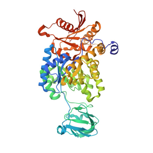
    The inclusion of novel small molecules in crystallization experiments has provided very encouraging results and this method is now emerging as a promising alternative strategy for crystallizing 'problematic' biological macromolecules. These small molecules have the ability to promote lattice formation through stabilizing intermolecular interactions in protein crystals. Here, the use of 1,3,6,8-pyrenetetrasulfonic acid (PTS), which provides a helpful intermolecular bridge between Leishmania mexicana PYK (LmPYK) macromolecules in the crystal, is reported, resulting in the rapid formation of a more stable crystal lattice at neutral pH and greatly improved X-ray diffraction results. The refined structure of the LmPYK-PTS complex revealed the negatively charged PTS molecule to be stacked between positively charged (surface-exposed) arginine side chains from neighbouring LmPYK molecules in the crystal lattice.


  • Organizational Affiliation
    • Structural Biochemistry Group, Institute of Structural and Molecular Biology, The University of Edinburgh, Michael Swann Building, The King's Buildings, Mayfield Road, Edinburgh EH9 3JR, Scotland.

Macromolecule Content 

  • Total Structure Weight: 110.01 kDa 
  • Atom Count: 8,214 
  • Modeled Residue Count: 984 
  • Deposited Residue Count: 998 
  • Unique protein chains: 1

Macromolecules

Find similar proteins by:|  3D Structure
Entity ID: 1
MoleculeChains  Sequence LengthOrganismDetailsImage
Pyruvate kinase
A, B
499Leishmania mexicanaMutation(s): 0 
Gene Names: PYK
EC: 2.7.1.40
UniProt
Find proteins for Q27686 (Leishmania mexicana)
Explore Q27686 
Go to UniProtKB:  Q27686
Entity Groups
Sequence Clusters30% Identity50% Identity70% Identity90% Identity95% Identity100% Identity
UniProt GroupQ27686
Sequence Annotations
Expand
Reference Sequence

Experimental Data & Validation

Experimental Data

  • Method: X-RAY DIFFRACTION
  • Resolution: 2.10 Å
  • R-Value Free:  0.239 (Depositor), 0.232 (DCC) 
  • R-Value Work:  0.186 (Depositor) 
  • R-Value Observed: 0.188 (Depositor) 
Space Group: I 2 2 2
Unit Cell:
Length ( Å )Angle ( ˚ )
a = 122.86α = 90
b = 129.86β = 90
c = 165.2γ = 90
Software Package:
Software NamePurpose
HKL-2000data collection
PHASERphasing
REFMACrefinement
DENZOdata reduction
SCALAdata scaling

Structure Validation

View Full Validation Report


Ligand Structure Quality Assessment 


Entry History 

Deposition Data

Revision History  (Full details and data files)

  • Version 1.0: 2010-02-09
    Type: Initial release
  • Version 1.1: 2011-07-13
    Changes: Version format compliance
  • Version 1.2: 2012-03-28
    Changes: Database references
  • Version 1.3: 2023-09-06
    Changes: Data collection, Database references, Derived calculations, Refinement description