3KNT | pdb_00003knt

Crystal structure of Methanocaldococcus jannaschii 8-oxoguanine glycosylase/lyase in complex with 15mer DNA containing 8-oxoguanine


Experimental Data Snapshot

  • Method: X-RAY DIFFRACTION
  • Resolution: 2.70 Å
  • R-Value Free: 
    0.225 (Depositor), 0.217 (DCC) 
  • R-Value Work: 
    0.188 (Depositor), 0.187 (DCC) 
  • R-Value Observed: 
    0.190 (Depositor) 

Starting Model: experimental
View more details

wwPDB Validation 3D Report Full Report

Validation slider image for 3KNT

This is version 1.4 of the entry. See complete history

Literature

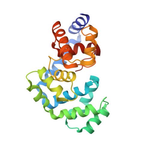
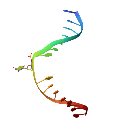
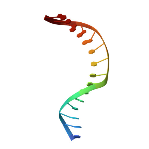
The C-terminal Lysine of Ogg2 DNA Glycosylases is a Major Molecular Determinant for Guanine/8-Oxoguanine Distinction.

Faucher, F.Wallace, S.S.Doublie, S.

(2010) J Mol Biology 397: 46-56

  • DOI: https://doi.org/10.1016/j.jmb.2010.01.024
  • Primary Citation Related Structures: 
    3KNT

  • PubMed Abstract: 

    7,8-Dihydro-8-oxoguanine (8-oxoG) is a major oxidative lesion found in DNA. The 8-oxoguanine DNA glycosylases (Ogg) responsible for the removal of 8-oxoG are divided into three families Ogg1, Ogg2 and AGOG. The Ogg2 members are devoid of the recognition loop used by Ogg1 to discriminate between 8-oxoG and guanine and it was unclear until recently how Ogg2 enzymes recognize the oxidized base. We present here the first crystallographic structure of an Ogg2 member, Methanocaldococcus janischii Ogg, in complex with a DNA duplex containing the 8-oxoG lesion. This structure highlights the crucial role of the C-terminal lysine, strictly conserved in Ogg2, in the recognition of 8-oxoG. The structure also reveals that Ogg2 undergoes a conformational change upon DNA binding similar to that observed in Ogg1 glycosylases. Furthermore, this work provides a structural rationale for the lack of opposite base specificity in this family of enzymes.


  • Organizational Affiliation
    • Department of Microbiology and Molecular Genetics, The Markey Center for Molecular Genetics, University of Vermont, Stafford Hall, 95 Carrigan Drive, Burlington, VT 05405-0068, USA.

Macromolecule Content 

  • Total Structure Weight: 136.35 kDa 
  • Atom Count: 9,326 
  • Modeled Residue Count: 938 
  • Deposited Residue Count: 948 
  • Unique protein chains: 1
  • Unique nucleic acid chains: 2

Macromolecules


Find similar proteins by:|  3D Structure
Entity ID: 1
MoleculeChains  Sequence LengthOrganismDetailsImage
N-glycosylase/DNA lyase
A, B, C, D
207Methanocaldococcus jannaschiiMutation(s): 1 
Gene Names: 1451601MJ0724ogg
EC: 3.2.2 (PDB Primary Data), 4.2.99.18 (PDB Primary Data)
UniProt
Find proteins for Q58134 (Methanocaldococcus jannaschii (strain ATCC 43067 / DSM 2661 / JAL-1 / JCM 10045 / NBRC 100440))
Explore Q58134 
Go to UniProtKB:  Q58134
Entity Groups
Sequence Clusters30% Identity50% Identity70% Identity90% Identity95% Identity100% Identity
UniProt GroupQ58134
Sequence Annotations
Expand
Reference Sequence
Find similar nucleic acids by:  Sequence
Entity ID: 2
MoleculeChains LengthOrganismImage
5'-D(*AP*CP*GP*TP*CP*CP*AP*(8OG)P*GP*TP*CP*TP*AP*CP*C)-3'
E, G, I, K
15N/A
Sequence Annotations
Expand
Reference Sequence
Find similar nucleic acids by:  Sequence
Entity ID: 3
MoleculeChains LengthOrganismImage
5'-D(*TP*GP*GP*TP*AP*GP*AP*CP*CP*TP*GP*GP*AP*CP*G)-3'
F, H, J, L
15N/A
Sequence Annotations
Expand
Reference Sequence

Experimental Data & Validation

Experimental Data

  • Method: X-RAY DIFFRACTION
  • Resolution: 2.70 Å
  • R-Value Free:  0.225 (Depositor), 0.217 (DCC) 
  • R-Value Work:  0.188 (Depositor), 0.187 (DCC) 
  • R-Value Observed: 0.190 (Depositor) 
Space Group: P 1 21 1
Unit Cell:
Length ( Å )Angle ( ˚ )
a = 54.8α = 90
b = 150.03β = 107.53
c = 90.27γ = 90
Software Package:
Software NamePurpose
Blu-Icedata collection
PHENIXmodel building
PHENIXrefinement
XDSdata reduction
XSCALEdata scaling
PHENIXphasing

Structure Validation

View Full Validation Report


Entry History 

Deposition Data

Revision History  (Full details and data files)

  • Version 1.0: 2010-02-02
    Type: Initial release
  • Version 1.1: 2011-07-13
    Changes: Version format compliance
  • Version 1.2: 2017-11-01
    Changes: Refinement description
  • Version 1.3: 2021-10-13
    Changes: Database references, Derived calculations
  • Version 1.4: 2023-09-06
    Changes: Data collection, Refinement description