3KL5

Structure Analysis of a Xylanase From Glycosyl Hydrolase Family Thirty: Carbohydrate Ligand Complexes Reveal this Family of Enzymes Unique Mechanism of Substrate Specificity and Recognition


Experimental Data Snapshot

  • Method: X-RAY DIFFRACTION
  • Resolution: 2.59 Å
  • R-Value Free: 0.290 
  • R-Value Work: 0.241 
  • R-Value Observed: 0.243 

wwPDB Validation   3D Report Full Report


This is version 2.1 of the entry. See complete history


Literature

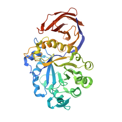
Ligand bound structures of a glycosyl hydrolase family 30 glucuronoxylan xylanohydrolase.

St John, F.J.Hurlbert, J.C.Rice, J.D.Preston, J.F.Pozharski, E.

(2011) J Mol Biol 407: 92-109

  • DOI: https://doi.org/10.1016/j.jmb.2011.01.010
  • Primary Citation of Related Structures:  
    3KL0, 3KL3, 3KL5

  • PubMed Abstract: 

    Xylanases of glycosyl hydrolase family 30 (GH30) have been shown to cleave β-1,4 linkages of 4-O-methylglucuronoxylan (MeGX(n)) as directed by the position along the xylan chain of an α-1,2-linked 4-O-methylglucuronate (MeGA) moiety. Complete hydrolysis of MeGX(n) by these enzymes results in singly substituted aldouronates having a 4-O-methylglucuronate moiety linked to a xylose penultimate from the reducing terminal xylose and some number of xylose residues toward the nonreducing terminus. This novel mode of action distinguishes GH30 xylanases from the more common xylanase families that cleave MeGX(n) in accessible regions. To help understand this unique biochemical function, we have determined the structure of XynC in its native and ligand-bound forms. XynC structure models derived from diffraction data of XynC crystal soaks with the simple sugar glucuronate (GA) and the tetrameric sugar 4-O-methyl-aldotetrauronate resulted in models containing GA and 4-O-methyl-aldotriuronate, respectively. Each is observed in two locations within XynC surface openings. Ligand coordination occurs within the XynC catalytic substrate binding cleft and on the structurally fused side β-domain, demonstrating a substrate targeting role for this putative carbohydrate binding module. Structural data reveal that GA acts as a primary functional appendage for recognition and hydrolysis of the MeGX(n) polymer by the protein. This work compares the structure of XynC with a previously reported homologous enzyme, XynA, from Erwinia chrysanthemi and analyzes the ligand binding sites. Our results identify the molecular interactions that define the unique function of XynC and homologous GH30 enzymes.


  • Organizational Affiliation

    Department of Pharmaceutical Sciences, University of Maryland School of Pharmacy, Baltimore, MD 21201, USA. fjstjohn@gmail.com


Macromolecules
Find similar proteins by:  (by identity cutoff)  |  3D Structure
Entity ID: 1
MoleculeChains Sequence LengthOrganismDetailsImage
Glucuronoxylanase xynC
A, B, C, D
401Bacillus subtilisMutation(s): 0 
Gene Names: BSU18150xynCynfF
EC: 3.2.1.136
UniProt
Find proteins for Q45070 (Bacillus subtilis (strain 168))
Explore Q45070 
Go to UniProtKB:  Q45070
Entity Groups  
Sequence Clusters30% Identity50% Identity70% Identity90% Identity95% Identity100% Identity
UniProt GroupQ45070
Sequence Annotations
Expand
  • Reference Sequence
Oligosaccharides

Help

Entity ID: 2
MoleculeChains Length2D Diagram Glycosylation3D Interactions
4-O-methyl-alpha-D-glucopyranuronic acid-(1-2)-beta-D-xylopyranose-(1-4)-beta-D-xylopyranose
E, F, G, H
3N/A
Glycosylation Resources
GlyTouCan:  G78055TN
GlyCosmos:  G78055TN
Experimental Data & Validation

Experimental Data

  • Method: X-RAY DIFFRACTION
  • Resolution: 2.59 Å
  • R-Value Free: 0.290 
  • R-Value Work: 0.241 
  • R-Value Observed: 0.243 
  • Space Group: P 21 21 2
Unit Cell:
Length ( Å )Angle ( ˚ )
a = 137.723α = 90
b = 194.008β = 90
c = 65.707γ = 90
Software Package:
Software NamePurpose
DENZOdata reduction
SCALEPACKdata scaling
PHASERphasing
REFMACrefinement
PDB_EXTRACTdata extraction
Blu-Icedata collection
Web-Icedata collection

Structure Validation

View Full Validation Report


Entry History 

Deposition Data

Revision History  (Full details and data files)

  • Version 1.0: 2010-12-08
    Type: Initial release
  • Version 1.1: 2011-07-13
    Changes: Version format compliance
  • Version 1.2: 2017-11-01
    Changes: Advisory, Refinement description
  • Version 2.0: 2020-07-29
    Type: Remediation
    Reason: Carbohydrate remediation
    Changes: Advisory, Atomic model, Data collection, Database references, Derived calculations, Structure summary
  • Version 2.1: 2023-09-06
    Changes: Data collection, Database references, Refinement description, Structure summary