3KH5

Crystal Structure of Protein MJ1225 from Methanocaldococcus jannaschii, a putative archaeal homolog of g-AMPK.


Experimental Data Snapshot

  • Method: X-RAY DIFFRACTION
  • Resolution: 2.10 Å
  • R-Value Free: 0.294 
  • R-Value Work: 0.228 
  • R-Value Observed: 0.231 

wwPDB Validation   3D Report Full Report


Ligand Structure Quality Assessment 


This is version 1.3 of the entry. See complete history


Literature

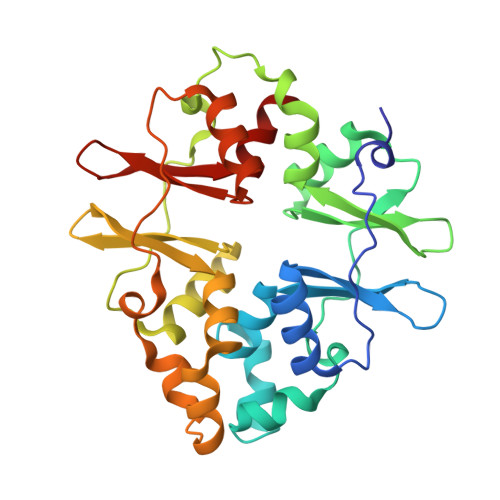
The crystal structure of protein MJ1225 from Methanocaldococcus jannaschii shows strong conservation of key structural features seen in the eukaryal gamma-AMPK.

Gomez-Garcia, I.Oyenarte, I.Martinez-Cruz, L.A.

(2010) J Mol Biol 65: 813-817

  • DOI: https://doi.org/10.1016/j.jmb.2010.03.045
  • Primary Citation of Related Structures:  
    3KH5, 3LFZ

  • PubMed Abstract: 

    In mammals, 5'-AMP-activated protein kinase (AMPK) is a heterotrimeric protein composed of a catalytic serine/threonine kinase subunit (alpha) and two regulatory subunits (beta and gamma). The gamma-subunit senses the intracellular energy status by competitively binding AMP and ATP and is thought to be responsible for allosteric regulation of the whole complex. We describe herein the crystal structure of protein MJ1225 from Methanocaldococcus jannaschii complexed to AMP, ADP, and ATP. Our data provide evidence of a strong conservation of the key functional features seen in the gamma-subunit of the eukaryotic AMPK, and more importantly, it reveals a novel AMP binding site, herein denoted as site E, which had not been previously described in cystathionine beta-synthase domains so far. Site E is located in a small cavity existing between the alpha-helices structurally equivalent to those disrupting the internal symmetry of each Bateman domain in gamma-AMPKs and shows striking similarities with a symmetry-related crevice of the mammalian enzyme that hosts the pathological mutation N488I.


  • Organizational Affiliation

    Structural Biology Unit, CIC bioGUNE, Parque Tecnológico de Bizkaia, Edificio 800, 48160 Derio, Bizkaia, Spain.


Macromolecules
Find similar proteins by:  (by identity cutoff)  |  3D Structure
Entity ID: 1
MoleculeChains Sequence LengthOrganismDetailsImage
protein MJ1225280Methanocaldococcus jannaschiiMutation(s): 0 
Gene Names: MJ1225
EC: 1.1.1.34
UniProt
Find proteins for Q58622 (Methanocaldococcus jannaschii (strain ATCC 43067 / DSM 2661 / JAL-1 / JCM 10045 / NBRC 100440))
Explore Q58622 
Go to UniProtKB:  Q58622
Entity Groups  
Sequence Clusters30% Identity50% Identity70% Identity90% Identity95% Identity100% Identity
UniProt GroupQ58622
Sequence Annotations
Expand
  • Reference Sequence
Experimental Data & Validation

Experimental Data

  • Method: X-RAY DIFFRACTION
  • Resolution: 2.10 Å
  • R-Value Free: 0.294 
  • R-Value Work: 0.228 
  • R-Value Observed: 0.231 
  • Space Group: H 3 2
Unit Cell:
Length ( Å )Angle ( ˚ )
a = 108.548α = 90
b = 108.548β = 90
c = 143.627γ = 120
Software Package:
Software NamePurpose
HKL-2000data collection
MOLREPphasing
REFMACrefinement
HKL-2000data reduction
HKL-2000data scaling

Structure Validation

View Full Validation Report


Ligand Structure Quality Assessment 


Entry History 

Deposition Data

Revision History  (Full details and data files)

  • Version 1.0: 2010-04-21
    Type: Initial release
  • Version 1.1: 2011-07-13
    Changes: Version format compliance
  • Version 1.2: 2024-02-21
    Changes: Data collection, Database references, Derived calculations
  • Version 1.3: 2024-04-03
    Changes: Refinement description