3KBP | pdb_00003kbp

KIDNEY BEAN PURPLE ACID PHOSPHATASE


Experimental Data Snapshot

  • Method: X-RAY DIFFRACTION
  • Resolution: 3.00 Å
  • R-Value Free: 
    0.220 (Depositor) 
  • R-Value Work: 
    0.189 (Depositor) 
  • R-Value Observed: 
    0.189 (Depositor) 

wwPDB Validation 3D Report Full Report

Validation slider image for 3KBP

This is version 1.4 of the entry. See complete history

Literature

Mechanism of Fe(III)-Zn(II) purple acid phosphatase based on crystal structures.

Klabunde, T.Strater, N.Frohlich, R.Witzel, H.Krebs, B.

(1996) J Mol Biology 259: 737-748

  • DOI: https://doi.org/10.1006/jmbi.1996.0354
  • Primary Citation Related Structures: 
    1KBP, 3KBP, 4KBP

  • PubMed Abstract: 

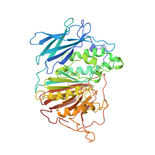
    Purple acid phosphatase is a widely distributed non-specific phosphomonoesterase. X-ray structures of the dimeric 111-kDa Fe(III)-Zn(II) kidney bean purple acid phosphatase (kbPAP) complexed with phosphate, the product of the reaction, and with tungstate, a strong inhibitor of the phosphatase activity, were determined at 2.7 and 3.0 angstroms resolution, respectively. Furthermore the resolution of the unligated enzyme, recently solved at 2.9 angstroms could be extended to 2.65 angstroms with completely new data. The binding of both oxoanions is not accompanied by larger conformational changes in the enzyme structure. Small movements with a maximal coordinate shift of 1 angstroms are only observed for the active site residues His295 and His296. In the inhibitor complex as well as in the product complex, the oxoanion binds in a bidentate bridging mode to the two metal ions, replacing two of the presumed solvent ligands present in the unligated enzyme form. As also proposed for the unligated structure a bridging hydroxide ion completes the coordination spheres of both metal ions to octahedral arrangements. All three structures reported herein support a mechanism of phosphate ester hydrolysis involving interaction of the substrate with Zn(II) followed by a nucleophilic attack on the phosphorus by an Fe(III)-coordinated hydroxide ion. The negative charge evolving at the pentacoordinated transition state is probably stabilized by interactions with the divalent zinc and the imidazole groups of His202, His295, and His296, the latter protonating the leaving alcohol group.


  • Organizational Affiliation
    • Anorganisch-Chemisches Institut, Universität Münster, Germany.

Macromolecule Content 

  • Total Structure Weight: 207.12 kDa 
  • Atom Count: 14,284 
  • Modeled Residue Count: 1,696 
  • Deposited Residue Count: 1,728 
  • Unique protein chains: 1

Macromolecules

Find similar proteins by:|  3D Structure
Entity ID: 1
MoleculeChains  Sequence LengthOrganismDetailsImage
PURPLE ACID PHOSPHATASE
A, B, C, D
432Phaseolus vulgarisMutation(s): 0 
EC: 3.1.3.2
UniProt
Find proteins for P80366 (Phaseolus vulgaris)
Explore P80366 
Go to UniProtKB:  P80366
Entity Groups
Sequence Clusters30% Identity50% Identity70% Identity90% Identity95% Identity100% Identity
UniProt GroupP80366
Glycosylation
Glycosylation Sites: 5
Sequence Annotations
Expand
Reference Sequence

Small Molecules

Ligands 4 Unique
IDChains Name / Formula / InChI Key2D Diagram3D Interactions
WO4

Query on WO4


Download:Ideal Coordinates CCD File
BA [auth C],
JA [auth D],
L [auth A],
T [auth B]
TUNGSTATE(VI)ION
O4 W
PBYZMCDFOULPGH-UHFFFAOYSA-N
NAG

Query on NAG


Download:Ideal Coordinates CCD File
CA [auth D]
DA [auth D]
E [auth A]
EA [auth D]
F [auth A]
CA [auth D],
DA [auth D],
E [auth A],
EA [auth D],
F [auth A],
FA [auth D],
G [auth A],
GA [auth D],
H [auth A],
I [auth A],
M [auth B],
N [auth B],
O [auth B],
P [auth B],
Q [auth B],
U [auth C],
V [auth C],
W [auth C],
X [auth C],
Y [auth C]
2-acetamido-2-deoxy-beta-D-glucopyranose
C8 H15 N O6
OVRNDRQMDRJTHS-FMDGEEDCSA-N
ZN

Query on ZN


Download:Ideal Coordinates CCD File
AA [auth C],
IA [auth D],
K [auth A],
S [auth B]
ZINC ION
Zn
PTFCDOFLOPIGGS-UHFFFAOYSA-N
FE

Query on FE


Download:Ideal Coordinates CCD File
HA [auth D],
J [auth A],
R [auth B],
Z [auth C]
FE (III) ION
Fe
VTLYFUHAOXGGBS-UHFFFAOYSA-N

Experimental Data & Validation

Experimental Data

  • Method: X-RAY DIFFRACTION
  • Resolution: 3.00 Å
  • R-Value Free:  0.220 (Depositor) 
  • R-Value Work:  0.189 (Depositor) 
  • R-Value Observed: 0.189 (Depositor) 
Space Group: C 2 2 21
Unit Cell:
Length ( Å )Angle ( ˚ )
a = 132.7α = 90
b = 347.3β = 90
c = 128.7γ = 90
Software Package:
Software NamePurpose
DENZOdata reduction
X-PLORmodel building
X-PLORrefinement
X-PLORphasing

Structure Validation

View Full Validation Report


Entry History 

Deposition Data

Revision History  (Full details and data files)

  • Version 1.0: 1996-12-07
    Type: Initial release
  • Version 1.1: 2008-03-25
    Changes: Version format compliance
  • Version 1.2: 2011-07-13
    Changes: Non-polymer description, Version format compliance
  • Version 1.3: 2020-07-29
    Type: Remediation
    Reason: Carbohydrate remediation
    Changes: Data collection, Database references, Derived calculations, Structure summary
  • Version 1.4: 2024-10-30
    Changes: Data collection, Database references, Structure summary