3K8P

Structural basis for vesicle tethering by the Dsl1 complex


Experimental Data Snapshot

  • Method: X-RAY DIFFRACTION
  • Resolution: 2.60 Å
  • R-Value Free: 0.271 
  • R-Value Work: 0.205 
  • R-Value Observed: 0.208 

wwPDB Validation   3D Report Full Report


This is version 1.1 of the entry. See complete history


Literature

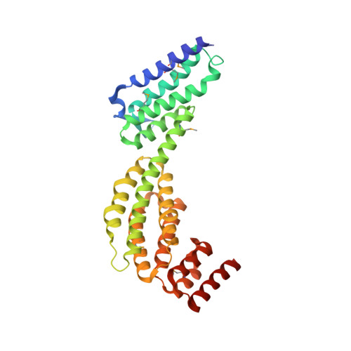
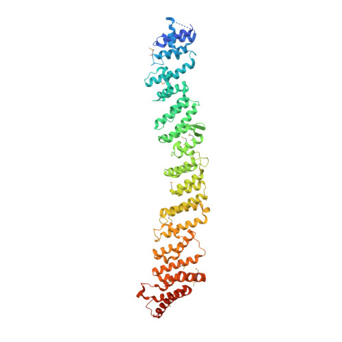
A structure-based mechanism for vesicle capture by the multisubunit tethering complex Dsl1.

Ren, Y.Yip, C.K.Tripathi, A.Huie, D.Jeffrey, P.D.Walz, T.Hughson, F.M.

(2009) Cell 139: 1119-1129

  • DOI: https://doi.org/10.1016/j.cell.2009.11.002
  • Primary Citation of Related Structures:  
    3K8P

  • PubMed Abstract: 
  • Vesicle trafficking requires membrane fusion, mediated by SNARE proteins, and upstream events that probably include "tethering," an initial long-range attachment between a vesicle and its target organelle. Among the factors proposed to mediate tethering are a set of multisubunit tethering complexes (MTCs) ...

    Vesicle trafficking requires membrane fusion, mediated by SNARE proteins, and upstream events that probably include "tethering," an initial long-range attachment between a vesicle and its target organelle. Among the factors proposed to mediate tethering are a set of multisubunit tethering complexes (MTCs). The Dsl1 complex, with only three subunits, is the simplest known MTC and is essential for the retrograde traffic of COPI-coated vesicles from the Golgi to the ER. To elucidate structural principles underlying MTC function, we have determined the structure of the Dsl1 complex, revealing a tower containing at its base the binding sites for two ER SNAREs and at its tip a flexible lasso for capturing vesicles. The Dsl1 complex binds to individual SNAREs via their N-terminal regulatory domains and also to assembled SNARE complexes; moreover, it is capable of accelerating SNARE complex assembly. Our results suggest that even the simplest MTC may be capable of orchestrating vesicle capture, uncoating, and fusion.


    Organizational Affiliation

    Department of Molecular Biology, Princeton University, Princeton, NJ 08544, USA.


Macromolecules
Find similar proteins by:  (by identity cutoff)  |  3D Structure
Entity ID: 1
MoleculeChainsSequence LengthOrganismDetailsImage
Dsl1A [auth C]357Kluyveromyces lactisMutation(s): 0 
Gene Names: KLLA0C02695g
UniProt
Find proteins for Q6CUS2 (Kluyveromyces lactis (strain ATCC 8585 / CBS 2359 / DSM 70799 / NBRC 1267 / NRRL Y-1140 / WM37))
Explore Q6CUS2 
Go to UniProtKB:  Q6CUS2
Entity Groups  
Sequence Clusters30% Identity50% Identity70% Identity90% Identity95% Identity100% Identity
UniProt GroupQ6CUS2
Protein Feature View
Expand
  • Reference Sequence
Find similar proteins by:  (by identity cutoff)  |  3D Structure
Entity ID: 2
MoleculeChainsSequence LengthOrganismDetailsImage
Protein transport protein SEC39B [auth D]709Saccharomyces cerevisiaeMutation(s): 0 
Gene Names: SEC39DSL3YLR440C
UniProt
Find proteins for Q12745 (Saccharomyces cerevisiae (strain ATCC 204508 / S288c))
Explore Q12745 
Go to UniProtKB:  Q12745
Entity Groups  
Sequence Clusters30% Identity50% Identity70% Identity90% Identity95% Identity100% Identity
UniProt GroupQ12745
Protein Feature View
Expand
  • Reference Sequence
Small Molecules
Modified Residues  1 Unique
IDChainsTypeFormula2D DiagramParent
MSE
Query on MSE
A [auth C]L-PEPTIDE LINKINGC5 H11 N O2 SeMET
Experimental Data & Validation

Experimental Data

  • Method: X-RAY DIFFRACTION
  • Resolution: 2.60 Å
  • R-Value Free: 0.271 
  • R-Value Work: 0.205 
  • R-Value Observed: 0.208 
  • Space Group: P 21 21 21
Unit Cell:
Length ( Å )Angle ( ˚ )
a = 72.02α = 90
b = 90.82β = 90
c = 213.88γ = 90
Software Package:
Software NamePurpose
CBASSdata collection
SHARPphasing
PHENIXrefinement
DENZOdata reduction
SCALEPACKdata scaling

Structure Validation

View Full Validation Report


Entry History 

Deposition Data

Revision History  (Full details and data files)

  • Version 1.0: 2009-11-10
    Type: Initial release
  • Version 1.1: 2011-07-13
    Changes: Version format compliance