3K6B

X-ray crystal structure of the E2 domain of APL-1 from C. elegans, in complex with sucrose octasulfate (SOS)


Experimental Data Snapshot

  • Method: X-RAY DIFFRACTION
  • Resolution: 2.80 Å
  • R-Value Free: 0.309 
  • R-Value Work: 0.262 
  • R-Value Observed: 0.262 

wwPDB Validation   3D Report Full Report


Ligand Structure Quality Assessment 


This is version 2.2 of the entry. See complete history


Literature

Structural characterization of the E2 domain of APL-1, a Caenorhabditis elegans homolog of human amyloid precursor protein, and its heparin binding site

Hoopes, J.T.Liu, X.Xu, X.Demeler, B.Folta-Stogniew, E.Li, C.Ha, Y.

(2010) J Biol Chem 285: 2165-2173

  • DOI: https://doi.org/10.1074/jbc.M109.018432
  • Primary Citation of Related Structures:  
    3K66, 3K6B

  • PubMed Abstract: 

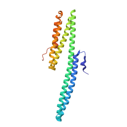
    The amyloid beta-peptide deposit found in the brain tissue of patients with Alzheimer disease is derived from a large heparin-binding protein precursor APP. The biological function of APP and its homologs is not precisely known. Here we report the x-ray structure of the E2 domain of APL-1, an APP homolog in Caenorhabditis elegans, and compare it to the human APP structure. We also describe the structure of APL-1 E2 in complex with sucrose octasulfate, a highly negatively charged disaccharide, which reveals an unexpected binding pocket between the two halves of E2. Based on the crystal structure, we are able to map, using site-directed mutagenesis, a surface groove on E2 to which heparin may bind. Our biochemical data also indicate that the affinity of E2 for heparin is influenced by pH: at pH 5, the binding appears to be much stronger than that at neutral pH. This property is likely caused by histidine residues in the vicinity of the mapped heparin binding site and could be important for the proposed adhesive function of APL-1.


  • Organizational Affiliation

    Department of Pharmacology, Yale School of Medicine, New Haven, CT 06520, USA.


Macromolecules
Find similar proteins by:  (by identity cutoff)  |  3D Structure
Entity ID: 1
MoleculeChains Sequence LengthOrganismDetailsImage
Beta-amyloid-like protein239Caenorhabditis elegansMutation(s): 0 
Gene Names: apl-1C42D8.8
UniProt
Find proteins for Q10651 (Caenorhabditis elegans)
Explore Q10651 
Go to UniProtKB:  Q10651
Entity Groups  
Sequence Clusters30% Identity50% Identity70% Identity90% Identity95% Identity100% Identity
UniProt GroupQ10651
Sequence Annotations
Expand
  • Reference Sequence
Small Molecules
Ligands 1 Unique
IDChains Name / Formula / InChI Key2D Diagram3D Interactions
GU4
Query on GU4

Download Ideal Coordinates CCD File 
B [auth A]2,3,4,6-tetra-O-sulfonato-alpha-D-glucopyranose
C6 H12 O18 S4
SPIXVQOXHJQNTH-DVKNGEFBSA-N
Experimental Data & Validation

Experimental Data

  • Method: X-RAY DIFFRACTION
  • Resolution: 2.80 Å
  • R-Value Free: 0.309 
  • R-Value Work: 0.262 
  • R-Value Observed: 0.262 
  • Space Group: P 32 2 1
Unit Cell:
Length ( Å )Angle ( ˚ )
a = 97.314α = 90
b = 97.314β = 90
c = 67.462γ = 120
Software Package:
Software NamePurpose
HKL-2000data collection
CNSrefinement
DENZOdata reduction
SCALEPACKdata scaling
CNSphasing

Structure Validation

View Full Validation Report


Ligand Structure Quality Assessment 


Entry History 

Deposition Data

Revision History  (Full details and data files)

  • Version 1.0: 2009-11-10
    Type: Initial release
  • Version 1.1: 2011-07-13
    Changes: Version format compliance
  • Version 2.0: 2020-04-22
    Changes: Atomic model, Data collection, Derived calculations, Non-polymer description, Structure summary
  • Version 2.1: 2020-07-29
    Type: Remediation
    Reason: Carbohydrate remediation
    Changes: Data collection, Derived calculations, Structure summary
  • Version 2.2: 2023-09-06
    Changes: Data collection, Database references, Refinement description, Structure summary