3JTH

Crystal structure of a transcriptional regulator HlyU from Vibrio vulnificus CMCP6


Experimental Data Snapshot

  • Method: X-RAY DIFFRACTION
  • Resolution: 2.00 Å
  • R-Value Free: 0.290 
  • R-Value Work: 0.231 
  • R-Value Observed: 0.242 

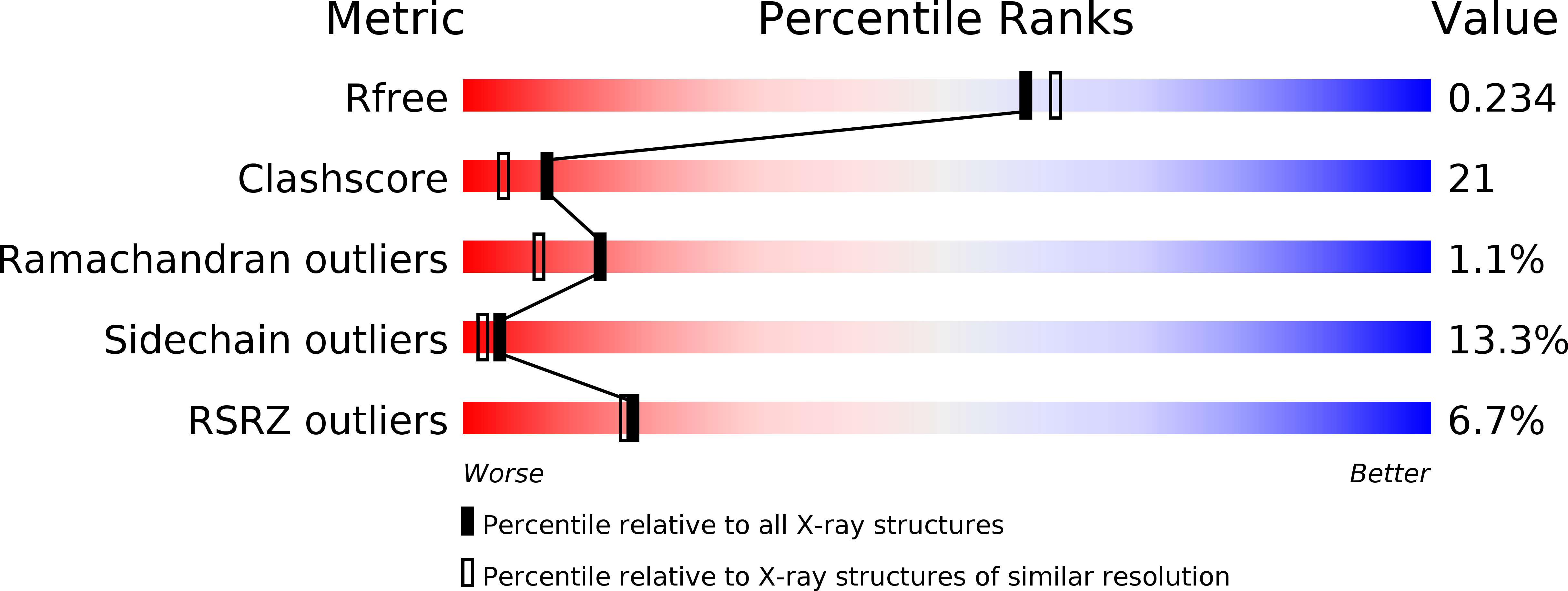
wwPDB Validation   3D Report Full Report


This is version 1.1 of the entry. See complete history


Literature

Crystal structure of the transcriptional activator HlyU from Vibrio vulnificus CMCP6.

Nishi, K.Lee, H.J.Park, S.Y.Bae, S.J.Lee, S.E.Adams, P.D.Rhee, J.H.Kim, J.S.

(2010) FEBS Lett 

  • DOI: https://doi.org/10.1016/j.febslet.2010.02.052
  • Primary Citation of Related Structures:  
    3JTH

  • PubMed Abstract: 

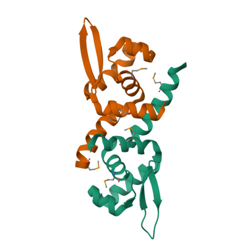
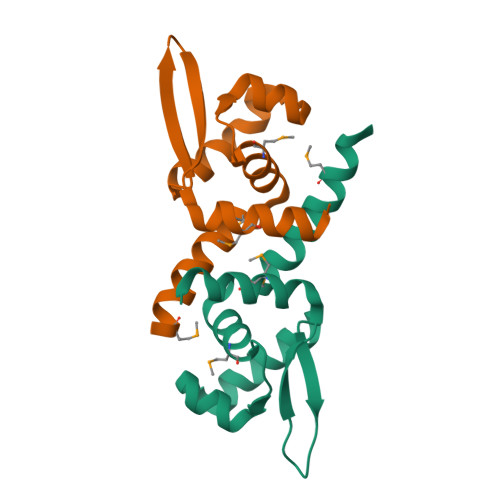
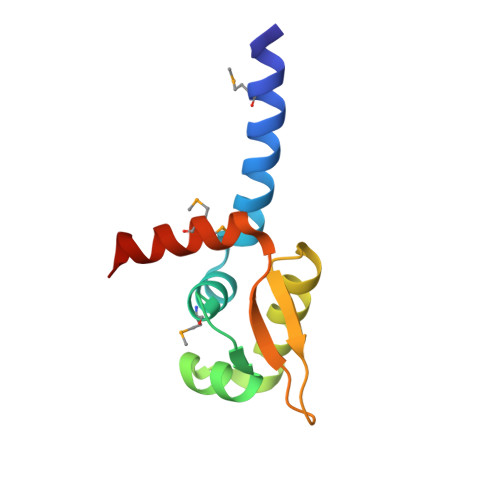
    HlyU is a transcription factor of the ArsR/SmtB family and activates the expression of the pathogenic Vibrio vulnificus RTX toxin. In contrast to the other metal-responding ArsR/SmtB proteins, HlyU does not sense metal ions. To provide its structural information, we elucidated the crystal structure of HlyU from V. vulnificus CMCP6 (HlyU_Vv). The monomeric HlyU_Vv architecture of five alpha-helices and two beta-strands, some of which constitute a typical DNA-binding winged helix-turn-helix (wHTH) motif, is very similar to that of other transcription regulators. Nonetheless, the homo-dimeric HlyU_Vv structure shows several different, three-dimensional features in the spatial position and the detailed dimeric interaction, which were not observed in the modeling study based on the same protein family and sequence similarity.


  • Organizational Affiliation

    Department of Chemistry, Chonnam National University, Gwangju, Republic of Korea.


Macromolecules
Find similar proteins by:  (by identity cutoff)  |  3D Structure
Entity ID: 1
MoleculeChains Sequence LengthOrganismDetailsImage
Transcription activator HlyU
A, B
98Vibrio vulnificus CMCP6Mutation(s): 0 
Gene Names: VV1_0512
Entity Groups  
Sequence Clusters30% Identity50% Identity70% Identity90% Identity95% Identity100% Identity
Sequence Annotations
Expand
  • Reference Sequence
Small Molecules
Modified Residues  1 Unique
IDChains TypeFormula2D DiagramParent
MSE
Query on MSE
A, B
L-PEPTIDE LINKINGC5 H11 N O2 SeMET
Experimental Data & Validation

Experimental Data

  • Method: X-RAY DIFFRACTION
  • Resolution: 2.00 Å
  • R-Value Free: 0.290 
  • R-Value Work: 0.231 
  • R-Value Observed: 0.242 
  • Space Group: P 1 21 1
Unit Cell:
Length ( Å )Angle ( ˚ )
a = 35.946α = 90
b = 52.79β = 103.92
c = 47.862γ = 90
Software Package:
Software NamePurpose
HKL-2000data collection
SOLVEphasing
CNSrefinement
HKL-2000data reduction
HKL-2000data scaling

Structure Validation

View Full Validation Report


Entry History 

Deposition Data

Revision History  (Full details and data files)

  • Version 1.0: 2010-03-16
    Type: Initial release
  • Version 1.1: 2011-07-13
    Changes: Version format compliance