Structure and filament dynamics of the pSK41 actin-like ParM protein: implications for plasmid DNA segregation.
Popp, D., Xu, W., Narita, A., Brzoska, A.J., Skurray, R.A., Firth, N., Goshdastider, U., Maeda, Y., Robinson, R.C., Schumacher, M.A.(2010) J Biological Chem 285: 10130-10140
- PubMed: 20106979 Search on PubMedSearch on PubMed Central
- DOI: https://doi.org/10.1074/jbc.M109.071613
- Primary Citation Related Structures:
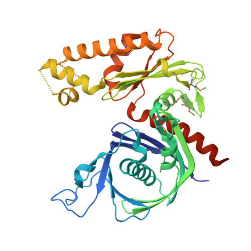
3JS6 - PubMed Abstract:
Type II plasmid partition systems utilize ParM NTPases in coordination with a centromere-binding protein called ParR to mediate accurate DNA segregation, a process critical for plasmid retention. The Staphylococcus aureus pSK41 plasmid is a medically important plasmid that confers resistance to multiple antibiotics, disinfectants, and antiseptics. In the first step of partition, the pSK41 ParR binds its DNA centromere to form a superhelical partition complex that recruits ParM, which then mediates plasmid separation. pSK41 ParM is homologous to R1 ParM, a known actin homologue, suggesting that it may also form filaments to drive partition. To gain insight into the partition function of ParM, we examined its ability to form filaments and determined the crystal structure of apoParM to 1.95 A. The structure shows that pSK41 ParM belongs to the actin/Hsp70 superfamily. Unexpectedly, however, pSK41 ParM shows the strongest structural homology to the archaeal actin-like protein Thermoplasma acidophilum Ta0583, rather than its functional homologue, R1 ParM. Consistent with this divergence, we find that regions shown to be involved in R1 ParM filament formation are not important in formation of pSK41 ParM polymers. These data are also consonant with our finding that pSK41 ParM forms 1-start 10/4 helices very different from the 37/17 symmetry of R1 ParM. The polymerization kinetics of pSK41 ParM also differed from that of R1 ParM. These results indicate that type II NTPases utilize different polymeric structures to drive plasmid segregation.
- ERATO "Actin Filament Dynamics" Project, Japan Science and Technology Corporation, c/o RIKEN, Harima Institute at Spring 8, 1-1-1 Kouto, Sayo, Hyogo 679-5148, Japan; Institute of Molecular and Cell Biology, 61 Biopolis Drive, Proteos 138673, Singapore. Electronic address: dpopp@imcb.A-star.edu.sg.
Organizational Affiliation: