3JQ7

Crystal structure of pteridine reductase 1 (PTR1) from Trypanosoma brucei in ternary complex with cofactor (NADP+) and inhibitor 6-phenylpteridine-2,4,7-triamine (DX2)


Experimental Data Snapshot

  • Method: X-RAY DIFFRACTION
  • Resolution: 1.80 Å
  • R-Value Free: 0.191 
  • R-Value Work: 0.152 
  • R-Value Observed: 0.154 

wwPDB Validation   3D Report Full Report


Ligand Structure Quality Assessment 


This is version 1.2 of the entry. See complete history


Literature

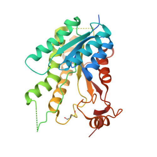
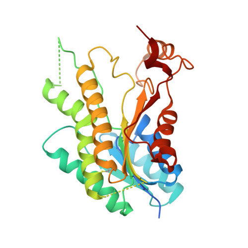
Structure-based design of pteridine reductase inhibitors targeting african sleeping sickness and the leishmaniases.

Tulloch, L.B.Martini, V.P.Iulek, J.Huggan, J.K.Lee, J.H.Gibson, C.L.Smith, T.K.Suckling, C.J.Hunter, W.N.

(2010) J Med Chem 53: 221-229

  • DOI: https://doi.org/10.1021/jm901059x
  • Primary Citation of Related Structures:  
    3BMC, 3BMN, 3BMO, 3BMQ, 3JQ6, 3JQ7, 3JQ8, 3JQ9, 3JQA, 3JQB, 3JQC, 3JQD, 3JQE, 3JQF, 3JQG

  • PubMed Abstract: 

    Pteridine reductase (PTR1) is a target for drug development against Trypanosoma and Leishmania species, parasites that cause serious tropical diseases and for which therapies are inadequate. We adopted a structure-based approach to the design of novel PTR1 inhibitors based on three molecular scaffolds. A series of compounds, most newly synthesized, were identified as inhibitors with PTR1-species specific properties explained by structural differences between the T. brucei and L. major enzymes. The most potent inhibitors target T. brucei PTR1, and two compounds displayed antiparasite activity against the bloodstream form of the parasite. PTR1 contributes to antifolate drug resistance by providing a molecular bypass of dihydrofolate reductase (DHFR) inhibition. Therefore, combining PTR1 and DHFR inhibitors might improve therapeutic efficacy. We tested two new compounds with known DHFR inhibitors. A synergistic effect was observed for one particular combination highlighting the potential of such an approach for treatment of African sleeping sickness.


  • Organizational Affiliation

    Division of Biological Chemistry and Drug Discovery, College of Life Sciences, University of Dundee, Dundee DD15EH, UK.


Macromolecules
Find similar proteins by:  (by identity cutoff)  |  3D Structure
Entity ID: 1
MoleculeChains Sequence LengthOrganismDetailsImage
Pteridine reductase 1
A, C, D
288Trypanosoma bruceiMutation(s): 0 
Gene Names: PTR1Tb927.8.2210
EC: 1.5.1.33
UniProt
Find proteins for Q581W1 (Trypanosoma brucei brucei (strain 927/4 GUTat10.1))
Explore Q581W1 
Go to UniProtKB:  Q581W1
Entity Groups  
Sequence Clusters30% Identity50% Identity70% Identity90% Identity95% Identity100% Identity
UniProt GroupQ581W1
Sequence Annotations
Expand
  • Reference Sequence
Find similar proteins by:  (by identity cutoff)  |  3D Structure
Entity ID: 2
MoleculeChains Sequence LengthOrganismDetailsImage
Pteridine reductase 1288Trypanosoma bruceiMutation(s): 0 
Gene Names: PTR1Tb927.8.2210
EC: 1.5.1.33
UniProt
Find proteins for Q581W1 (Trypanosoma brucei brucei (strain 927/4 GUTat10.1))
Explore Q581W1 
Go to UniProtKB:  Q581W1
Entity Groups  
Sequence Clusters30% Identity50% Identity70% Identity90% Identity95% Identity100% Identity
UniProt GroupQ581W1
Sequence Annotations
Expand
  • Reference Sequence
Small Molecules
Ligands 4 Unique
IDChains Name / Formula / InChI Key2D Diagram3D Interactions
NAP
Query on NAP

Download Ideal Coordinates CCD File 
E [auth A],
H [auth B],
L [auth C],
N [auth D]
NADP NICOTINAMIDE-ADENINE-DINUCLEOTIDE PHOSPHATE
C21 H28 N7 O17 P3
XJLXINKUBYWONI-NNYOXOHSSA-N
DX2
Query on DX2

Download Ideal Coordinates CCD File 
F [auth A],
I [auth B],
M [auth C],
O [auth D]
6-phenylpteridine-2,4,7-triamine
C12 H11 N7
FNYLWPVRPXGIIP-UHFFFAOYSA-N
DTT
Query on DTT

Download Ideal Coordinates CCD File 
J [auth B]2,3-DIHYDROXY-1,4-DITHIOBUTANE
C4 H10 O2 S2
VHJLVAABSRFDPM-IMJSIDKUSA-N
ACT
Query on ACT

Download Ideal Coordinates CCD File 
G [auth A],
K [auth B]
ACETATE ION
C2 H3 O2
QTBSBXVTEAMEQO-UHFFFAOYSA-M
Modified Residues  1 Unique
IDChains TypeFormula2D DiagramParent
CSX
Query on CSX
A, C, D
L-PEPTIDE LINKINGC3 H7 N O3 SCYS
Binding Affinity Annotations 
IDSourceBinding Affinity
DX2 Binding MOAD:  3JQ7 Ki: 1200 (nM) from 1 assay(s)
PDBBind:  3JQ7 Ki: 1200 (nM) from 1 assay(s)
Experimental Data & Validation

Experimental Data

  • Method: X-RAY DIFFRACTION
  • Resolution: 1.80 Å
  • R-Value Free: 0.191 
  • R-Value Work: 0.152 
  • R-Value Observed: 0.154 
  • Space Group: P 1 21 1
Unit Cell:
Length ( Å )Angle ( ˚ )
a = 74.538α = 90
b = 90.029β = 115.49
c = 82.359γ = 90
Software Package:
Software NamePurpose
MOSFLMdata reduction
SCALAdata scaling
REFMACrefinement
PDB_EXTRACTdata extraction
ADSCdata collection
MOLREPphasing

Structure Validation

View Full Validation Report


Ligand Structure Quality Assessment 


Entry History 

Deposition Data

Revision History  (Full details and data files)

  • Version 1.0: 2009-12-08
    Type: Initial release
  • Version 1.1: 2011-07-13
    Changes: Advisory, Refinement description, Version format compliance
  • Version 1.2: 2023-09-06
    Changes: Data collection, Database references, Derived calculations, Refinement description