3J5V

PhuZ201 filament


Experimental Data Snapshot

  • Method: ELECTRON MICROSCOPY
  • Resolution: 7.10 Å
  • Aggregation State: FILAMENT 
  • Reconstruction Method: HELICAL 

wwPDB Validation   3D Report Full Report


This is version 1.4 of the entry. See complete history


Literature

The Structure and Assembly Mechanism of a Novel Three-Stranded Tubulin Filament that Centers Phage DNA

Zehr, E.A.Kraemer, J.K.Erb, M.L.Coker, J.K.C.Montabana, E.A.Pogliano, J.Agard, D.A.

(2014) Structure 22: 1-10

  • DOI: https://doi.org/10.1016/j.str.2014.02.006
  • Primary Citation of Related Structures:  
    3J5V

  • PubMed Abstract: 

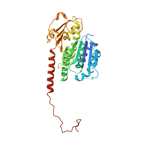
    Tubulins are a universally conserved protein superfamily that carry out diverse biological roles by assembling filaments with very different architectures. The underlying basis of this structural diversity is poorly understood. Here, we determine a 7.1 Å cryo-electron microscopy reconstruction of the bacteriophage-encoded PhuZ filament and provide molecular-level insight into its cooperative assembly mechanism. The PhuZ family of tubulins is required to actively center the phage within infected host cells, facilitating efficient phage replication. Our reconstruction and derived model reveal the first example of a three-stranded tubulin filament. We show that the elongated C-terminal tail simultaneously stabilizes both longitudinal and lateral interactions, which in turn define filament architecture. Identified interaction surfaces are conserved within the PhuZ family, and their mutagenesis compromises polymerization in vitro and in vivo. Combining kinetic modeling of PhuZ filament assembly and structural data, we suggest a common filament structure and assembly mechanism for the PhuZ family of tubulins.


  • Organizational Affiliation

    Department of Biochemistry and Biophysics and the Howard Hughes Medical Institute, University of California, San Francisco, San Francisco, CA 94158, USA.


Macromolecules
Find similar proteins by:  (by identity cutoff)  |  3D Structure
Entity ID: 1
MoleculeChains Sequence LengthOrganismDetailsImage
PhuZ201 subunitA [auth a]320Pseudomonas phage 201phi2-1Mutation(s): 0 
Gene Names: 201phi2-1p059
UniProt
Find proteins for B3FK34 (Pseudomonas phage 201phi2-1)
Explore B3FK34 
Go to UniProtKB:  B3FK34
Entity Groups  
Sequence Clusters30% Identity50% Identity70% Identity90% Identity95% Identity100% Identity
UniProt GroupB3FK34
Sequence Annotations
Expand
  • Reference Sequence
Small Molecules
Ligands 2 Unique
IDChains Name / Formula / InChI Key2D Diagram3D Interactions
GDP
Query on GDP

Download Ideal Coordinates CCD File 
B [auth a]GUANOSINE-5'-DIPHOSPHATE
C10 H15 N5 O11 P2
QGWNDRXFNXRZMB-UUOKFMHZSA-N
MG
Query on MG

Download Ideal Coordinates CCD File 
C [auth a]MAGNESIUM ION
Mg
JLVVSXFLKOJNIY-UHFFFAOYSA-N
Experimental Data & Validation

Experimental Data

  • Method: ELECTRON MICROSCOPY
  • Resolution: 7.10 Å
  • Aggregation State: FILAMENT 
  • Reconstruction Method: HELICAL 
EM Software:
TaskSoftware PackageVersion
RECONSTRUCTIONSPIDER

Structure Validation

View Full Validation Report


Entry History 

Deposition Data

  • Released Date: 2014-03-26 
  • Deposition Author(s): Zehr, E.A.

Revision History  (Full details and data files)

  • Version 1.0: 2014-03-26
    Type: Initial release
  • Version 1.1: 2014-04-09
    Changes: Other
  • Version 1.2: 2014-04-23
    Changes: Refinement description
  • Version 1.3: 2018-07-18
    Changes: Author supporting evidence, Data collection
  • Version 1.4: 2024-02-21
    Changes: Data collection, Database references, Derived calculations, Refinement description