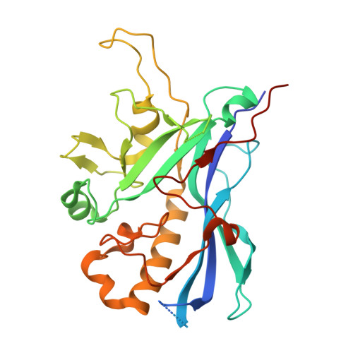
3J0I

Fitting of the phiKZ gp29PR structure into the cryo-EM density map of the phiKZ polysheath


Experimental Data Snapshot

  • Method: ELECTRON MICROSCOPY
  • Resolution: 19.0 Å
  • Aggregation State: PARTICLE 
  • Reconstruction Method: SINGLE PARTICLE 

wwPDB Validation   3D Report Full Report


This is version 1.6 of the entry. See complete history


Literature

Structural conservation of the myoviridae phage tail sheath protein fold.

Aksyuk, A.A.Kurochkina, L.P.Fokine, A.Forouhar, F.Mesyanzhinov, V.V.Tong, L.Rossmann, M.G.

(2011) Structure 19: 1885-1894

  • DOI: https://doi.org/10.1016/j.str.2011.09.012
  • Primary Citation of Related Structures:  
    3J0H, 3J0I, 3SPE

  • PubMed Abstract: 

    Bacteriophage phiKZ is a giant phage that infects Pseudomonas aeruginosa, a human pathogen. The phiKZ virion consists of a 1450 Å diameter icosahedral head and a 2000 Å-long contractile tail. The structure of the whole virus was previously reported, showing that its tail organization in the extended state is similar to the well-studied Myovirus bacteriophage T4 tail. The crystal structure of a tail sheath protein fragment of phiKZ was determined to 2.4 Å resolution. Furthermore, crystal structures of two prophage tail sheath proteins were determined to 1.9 and 3.3 Å resolution. Despite low sequence identity between these proteins, all of these structures have a similar fold. The crystal structure of the phiKZ tail sheath protein has been fitted into cryo-electron-microscopy reconstructions of the extended tail sheath and of a polysheath. The structural rearrangement of the phiKZ tail sheath contraction was found to be similar to that of phage T4.


  • Organizational Affiliation

    Department of Biological Sciences, Purdue University, West Lafayette, IN 47907-2032, USA.


Macromolecules
Find similar proteins by:  (by identity cutoff)  |  3D Structure
Entity ID: 1
MoleculeChains Sequence LengthOrganismDetailsImage
PHIKZ029
A, B, C, D, E
A, B, C, D, E, F
295Phikzvirus phiKZMutation(s): 0 
UniProt
Find proteins for Q8SDD3 (Pseudomonas phage phiKZ)
Explore Q8SDD3 
Go to UniProtKB:  Q8SDD3
Entity Groups  
Sequence Clusters30% Identity50% Identity70% Identity90% Identity95% Identity100% Identity
UniProt GroupQ8SDD3
Sequence Annotations
Expand
  • Reference Sequence
Experimental Data & Validation

Experimental Data

  • Method: ELECTRON MICROSCOPY
  • Resolution: 19.0 Å
  • Aggregation State: PARTICLE 
  • Reconstruction Method: SINGLE PARTICLE 
EM Software:
TaskSoftware PackageVersion
RECONSTRUCTIONSPIDER

Structure Validation

View Full Validation Report


Entry History 

Deposition Data

Revision History  (Full details and data files)

  • Version 1.0: 2011-12-14
    Type: Initial release
  • Version 1.1: 2012-03-28
    Changes: Database references
  • Version 1.2: 2012-07-25
    Changes: Other
  • Version 1.3: 2013-03-20
    Changes: Other
  • Version 1.4: 2014-07-23
    Changes: Other
  • Version 1.5: 2018-07-18
    Changes: Data collection
  • Version 1.6: 2024-02-21
    Changes: Data collection, Database references, Refinement description