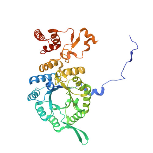
3IVT

Homocitrate Synthase Lys4 bound to 2-OG


Experimental Data Snapshot

  • Method: X-RAY DIFFRACTION
  • Resolution: 2.67 Å
  • R-Value Free: 0.206 
  • R-Value Work: 0.165 
  • R-Value Observed: 0.167 

Starting Model: other
View more details

wwPDB Validation   3D Report Full Report


This is version 1.5 of the entry. See complete history


Literature

Crystal structure and functional analysis of homocitrate synthase, an essential enzyme in lysine biosynthesis.

Bulfer, S.L.Scott, E.M.Couture, J.F.Pillus, L.Trievel, R.C.

(2009) J Biol Chem 284: 35769-35780

  • DOI: https://doi.org/10.1074/jbc.M109.046821
  • Primary Citation of Related Structures:  
    3IVS, 3IVT, 3IVU

  • PubMed Abstract: 

    Homocitrate synthase (HCS) catalyzes the first and committed step in lysine biosynthesis in many fungi and certain Archaea and is a potential target for antifungal drugs. Here we report the crystal structure of the HCS apoenzyme from Schizosaccharomyces pombe and two distinct structures of the enzyme in complex with the substrate 2-oxoglutarate (2-OG). The structures reveal that HCS forms an intertwined homodimer stabilized by domain-swapping between the N- and C-terminal domains of each monomer. The N-terminal catalytic domain is composed of a TIM barrel fold in which 2-OG binds via hydrogen bonds and coordination to the active site divalent metal ion, whereas the C-terminal domain is composed of mixed alpha/beta topology. In the structures of the HCS apoenzyme and one of the 2-OG binary complexes, a lid motif from the C-terminal domain occludes the entrance to the active site of the neighboring monomer, whereas in the second 2-OG complex the lid is disordered, suggesting that it regulates substrate access to the active site through its apparent flexibility. Mutations of the active site residues involved in 2-OG binding or implicated in acid-base catalysis impair or abolish activity in vitro and in vivo. Together, these results yield new insights into the structure and catalytic mechanism of HCSs and furnish a platform for developing HCS-selective inhibitors.


  • Organizational Affiliation

    Department of Biological Chemistry, University of Michigan, Ann Arbor, Michigan 48109, USA.


Macromolecules
Find similar proteins by:  (by identity cutoff)  |  3D Structure
Entity ID: 1
MoleculeChains Sequence LengthOrganismDetailsImage
Homocitrate synthase, mitochondrial
A, B
423Schizosaccharomyces pombeMutation(s): 0 
Gene Names: lys4SPBC1105.02c
EC: 2.3.3.14
UniProt
Find proteins for Q9Y823 (Schizosaccharomyces pombe (strain 972 / ATCC 24843))
Explore Q9Y823 
Go to UniProtKB:  Q9Y823
Entity Groups  
Sequence Clusters30% Identity50% Identity70% Identity90% Identity95% Identity100% Identity
UniProt GroupQ9Y823
Sequence Annotations
Expand
  • Reference Sequence
Experimental Data & Validation

Experimental Data

  • Method: X-RAY DIFFRACTION
  • Resolution: 2.67 Å
  • R-Value Free: 0.206 
  • R-Value Work: 0.165 
  • R-Value Observed: 0.167 
  • Space Group: P 62
Unit Cell:
Length ( Å )Angle ( ˚ )
a = 133.731α = 90
b = 133.731β = 90
c = 125.904γ = 120
Software Package:
Software NamePurpose
DENZOdata reduction
SCALEPACKdata scaling
MOLREPphasing
REFMACrefinement
PDB_EXTRACTdata extraction
HKL-2000data collection
HKL-2000data reduction
HKL-2000data scaling

Structure Validation

View Full Validation Report


Entry History 

Deposition Data

Revision History  (Full details and data files)

  • Version 1.0: 2009-09-22
    Type: Initial release
  • Version 1.1: 2011-07-13
    Changes: Advisory, Version format compliance
  • Version 1.2: 2017-11-01
    Changes: Refinement description
  • Version 1.3: 2018-01-24
    Changes: Structure summary
  • Version 1.4: 2024-02-21
    Changes: Data collection, Database references, Derived calculations
  • Version 1.5: 2024-04-03
    Changes: Refinement description