Crystal structure of prolyl 4-hydroxylase from Bacillus anthracis.
Culpepper, M.A., Scott, E.E., Limburg, J.(2010) Biochemistry 49: 124-133
- PubMed: 19947658 Search on PubMedSearch on PubMed Central
- DOI: https://doi.org/10.1021/bi901771z
- Primary Citation Related Structures:
3ITQ - PubMed Abstract:
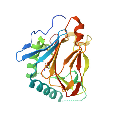
Prolyl 4-hydroxylases (P4H) catalyze the post-translational hydroxylation of proline residues and play a role in collagen production, hypoxia response, and cell wall development. P4Hs belong to the group of Fe(II)/alphaKG oxygenases and require Fe(II), alpha-ketoglutarate (alphaKG), and O(2) for activity. We report the 1.40 A structure of a P4H from Bacillus anthracis, the causative agent of anthrax, whose immunodominant exosporium protein BclA contains collagen-like repeat sequences. The structure reveals the double-stranded beta-helix core fold characteristic of Fe(II)/alphaKG oxygenases. This fold positions Fe-binding and alphaKG-binding residues in what is expected to be catalytically competent orientations and is consistent with proline peptide substrate binding at the active site mouth. Comparisons of the anthrax P4H structure with Cr P4H-1 structures reveal similarities in a peptide surface groove. However, sequence and structural comparisons suggest differences in conformation of adjacent loops may change the interaction with peptide substrates. These differences may be the basis of a substantial disparity between the K(M) values for the Cr P4H-1 compared to the anthrax and human P4H enzymes. Additionally, while previous structures of P4H enzymes are monomers, B. anthracis P4H forms an alpha(2) homodimer and suggests residues important for interactions between the alpha(2) subunits of alpha(2)beta(2) human collagen P4H. Thus, the anthrax P4H structure provides insight into the structure and function of the alpha-subunit of human P4H, which may aid in the development of selective inhibitors of the human P4H enzyme involved in fibrotic disease.
- Department of Chemistry, University of Kansas, 1251 Wescoe Hall Drive, Lawrence, Kansas 66045, USA.
Organizational Affiliation: