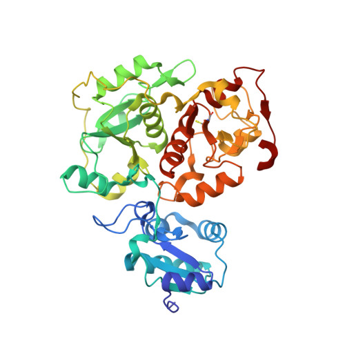
3IPO

Crystal structure of YnjE


Experimental Data Snapshot

  • Method: X-RAY DIFFRACTION
  • Resolution: 2.40 Å
  • R-Value Free: 0.211 
  • R-Value Work: 0.159 
  • R-Value Observed: 0.161 

wwPDB Validation   3D Report Full Report


This is version 1.1 of the entry. See complete history


Literature

Crystal structure of YnjE from Escherichia coli, a sulfurtransferase with three rhodanese domains.

Hanzelmann, P.Dahl, J.U.Kuper, J.Urban, A.Muller-Theissen, U.Leimkuhler, S.Schindelin, H.

(2009) Protein Sci 18: 2480-2491

  • DOI: https://doi.org/10.1002/pro.260
  • Primary Citation of Related Structures:  
    3IPO, 3IPP

  • PubMed Abstract: 

    Rhodaneses/sulfurtransferases are ubiquitous enzymes that catalyze the transfer of sulfane sulfur from a donor molecule to a thiophilic acceptor via an active site cysteine that is modified to a persulfide during the reaction. Here, we present the first crystal structure of a triple-domain rhodanese-like protein, namely YnjE from Escherichia coli, in two states where its active site cysteine is either unmodified or present as a persulfide. Compared to well-characterized tandem domain rhodaneses, which are composed of one inactive and one active domain, YnjE contains an extra N-terminal inactive rhodanese-like domain. Phylogenetic analysis reveals that YnjE triple-domain homologs can be found in a variety of other gamma-proteobacteria, in addition, some single-, tandem-, four and even six-domain variants exist. All YnjE rhodaneses are characterized by a highly conserved active site loop (CGTGWR) and evolved independently from other rhodaneses, thus forming their own subfamily. On the basis of structural comparisons with other rhodaneses and kinetic studies, YnjE, which is more similar to thiosulfate:cyanide sulfurtransferases than to 3-mercaptopyruvate:cyanide sulfurtransferases, has a different substrate specificity that depends not only on the composition of the active site loop with the catalytic cysteine at the first position but also on the surrounding residues. In vitro YnjE can be efficiently persulfurated by the cysteine desulfurase IscS. The catalytic site is located within an elongated cleft, formed by the central and C-terminal domain and is lined by bulky hydrophobic residues with the catalytic active cysteine largely shielded from the solvent.


  • Organizational Affiliation

    Rudolf Virchow Center for Experimental Biomedicine, University of Würzburg, 97080 Würzburg, Germany. petra.haenzelmann@virchow.uni-wuerzburg.de


Macromolecules
Find similar proteins by:  (by identity cutoff)  |  3D Structure
Entity ID: 1
MoleculeChains Sequence LengthOrganismDetailsImage
Putative thiosulfate sulfurtransferase ynjE
A, B
416Escherichia coli K-12Mutation(s): 0 
Gene Names: b1757JW5287ynjE
EC: 2.8.1.1
UniProt
Find proteins for P78067 (Escherichia coli (strain K12))
Explore P78067 
Go to UniProtKB:  P78067
Entity Groups  
Sequence Clusters30% Identity50% Identity70% Identity90% Identity95% Identity100% Identity
UniProt GroupP78067
Sequence Annotations
Expand
  • Reference Sequence
Small Molecules
Ligands 5 Unique
IDChains Name / Formula / InChI Key2D Diagram3D Interactions
PE4
Query on PE4

Download Ideal Coordinates CCD File 
E [auth B]2-{2-[2-(2-{2-[2-(2-ETHOXY-ETHOXY)-ETHOXY]-ETHOXY}-ETHOXY)-ETHOXY]-ETHOXY}-ETHANOL
C16 H34 O8
PJWQOENWHPEPKI-UHFFFAOYSA-N
PG0
Query on PG0

Download Ideal Coordinates CCD File 
G [auth B],
H [auth B]
2-(2-METHOXYETHOXY)ETHANOL
C5 H12 O3
SBASXUCJHJRPEV-UHFFFAOYSA-N
PO4
Query on PO4

Download Ideal Coordinates CCD File 
C [auth A],
I [auth B]
PHOSPHATE ION
O4 P
NBIIXXVUZAFLBC-UHFFFAOYSA-K
GOL
Query on GOL

Download Ideal Coordinates CCD File 
F [auth B]GLYCEROL
C3 H8 O3
PEDCQBHIVMGVHV-UHFFFAOYSA-N
NA
Query on NA

Download Ideal Coordinates CCD File 
D [auth A],
J [auth B]
SODIUM ION
Na
FKNQFGJONOIPTF-UHFFFAOYSA-N
Modified Residues  1 Unique
IDChains TypeFormula2D DiagramParent
CSS
Query on CSS
A, B
L-PEPTIDE LINKINGC3 H7 N O2 S2CYS
Experimental Data & Validation

Experimental Data

  • Method: X-RAY DIFFRACTION
  • Resolution: 2.40 Å
  • R-Value Free: 0.211 
  • R-Value Work: 0.159 
  • R-Value Observed: 0.161 
  • Space Group: P 63 2 2
Unit Cell:
Length ( Å )Angle ( ˚ )
a = 160.949α = 90
b = 160.949β = 90
c = 173.992γ = 120
Software Package:
Software NamePurpose
SCALAdata scaling
REFMACrefinement
PDB_EXTRACTdata extraction
MxCuBEdata collection
MOSFLMdata reduction
SHARPphasing

Structure Validation

View Full Validation Report


Entry History 

Deposition Data

Revision History  (Full details and data files)

  • Version 1.0: 2009-12-08
    Type: Initial release
  • Version 1.1: 2011-07-13
    Changes: Advisory, Refinement description, Version format compliance