3IO5

Crystal Structure of a dimeric form of the uvsX Recombinase core domain from Enterobacteria Phage T4


Experimental Data Snapshot

  • Method: X-RAY DIFFRACTION
  • Resolution: 2.40 Å
  • R-Value Free: 0.248 
  • R-Value Work: 0.210 
  • R-Value Observed: 0.212 

wwPDB Validation   3D Report Full Report


This is version 1.3 of the entry. See complete history


Literature

Crystal Structure of the Phage T4 Recombinase UvsX and Its Functional Interaction with the T4 SF2 Helicase UvsW.

Gajewski, S.Webb, M.R.Galkin, V.Egelman, E.H.Kreuzer, K.N.White, S.W.

(2011) J Mol Biol 405: 65-76

  • DOI: https://doi.org/10.1016/j.jmb.2010.10.004
  • Primary Citation of Related Structures:  
    3IO5

  • PubMed Abstract: 

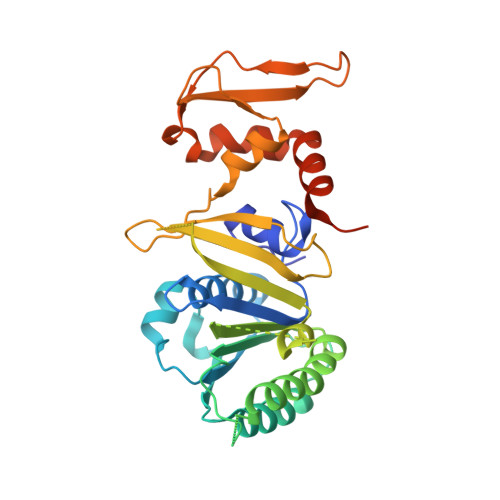
    Bacteriophage T4 provides an important model system for studying the mechanism of homologous recombination. We have determined the crystal structure of the T4 UvsX recombinase, and the overall architecture and fold closely resemble those of RecA, including a highly conserved ATP binding site. Based on this new structure, we reanalyzed electron microscopy reconstructions of UvsX-DNA filaments and docked the UvsX crystal structure into two different filament forms: a compressed filament generated in the presence of ADP and an elongated filament generated in the presence of ATP and aluminum fluoride. In these reconstructions, the ATP binding site sits at the protomer interface, as in the RecA filament crystal structure. However, the environment of the ATP binding site is altered in the two filament reconstructions, suggesting that nucleotide cannot be as easily accommodated at the protomer interface of the compressed filament. Finally, we show that the phage helicase UvsW completes the UvsX-promoted strand-exchange reaction, allowing the generation of a simple nicked circular product rather than complex networks of partially exchanged substrates.


  • Organizational Affiliation

    Department of Structural Biology, St. Jude Children's Research Hospital, Memphis, TN 38105, USA.


Macromolecules
Find similar proteins by:  (by identity cutoff)  |  3D Structure
Entity ID: 1
MoleculeChains Sequence LengthOrganismDetailsImage
Recombination and repair protein
A, B
333Tequatrovirus T4Mutation(s): 0 
Gene Names: fdsAUVSX
UniProt
Find proteins for P04529 (Enterobacteria phage T4)
Explore P04529 
Go to UniProtKB:  P04529
Entity Groups  
Sequence Clusters30% Identity50% Identity70% Identity90% Identity95% Identity100% Identity
UniProt GroupP04529
Sequence Annotations
Expand
  • Reference Sequence
Experimental Data & Validation

Experimental Data

  • Method: X-RAY DIFFRACTION
  • Resolution: 2.40 Å
  • R-Value Free: 0.248 
  • R-Value Work: 0.210 
  • R-Value Observed: 0.212 
  • Space Group: P 61
Unit Cell:
Length ( Å )Angle ( ˚ )
a = 95.974α = 90
b = 95.974β = 90
c = 131.274γ = 120
Software Package:
Software NamePurpose
DENZOdata reduction
SCALEPACKdata scaling
REFMACrefinement
PDB_EXTRACTdata extraction
MAR345dtbdata collection
HKL-2000data reduction
HKL-2000data scaling
PHENIXphasing

Structure Validation

View Full Validation Report


Entry History 

Deposition Data

  • Released Date: 2010-08-25 
  • Deposition Author(s): Gajewski, S.

Revision History  (Full details and data files)

  • Version 1.0: 2010-08-25
    Type: Initial release
  • Version 1.1: 2011-07-13
    Changes: Version format compliance
  • Version 1.2: 2017-11-01
    Changes: Refinement description
  • Version 1.3: 2024-02-21
    Changes: Data collection, Database references, Derived calculations