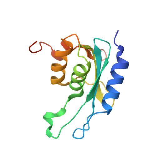
Structural and functional analyses of Mycobacterium tuberculosis Rv3315c-encoded metal-dependent homotetrameric cytidine deaminase.
Sanchez-Quitian, Z.A., Schneider, C.Z., Ducati, R.G., de Azevedo, W.F., Bloch, C., Basso, L.A., Santos, D.S.(2010) J Struct Biol 169: 413-423
- PubMed: 20035876
- DOI: https://doi.org/10.1016/j.jsb.2009.12.019
- Primary Citation of Related Structures:
3IJF - PubMed Abstract:
The emergence of drug-resistant strains of Mycobacterium tuberculosis, the causative agent of tuberculosis, has exacerbated the treatment and control of this disease. Cytidine deaminase (CDA) is a pyrimidine salvage pathway enzyme that recycles cytidine and 2'-deoxycytidine for uridine and 2'-deoxyuridine synthesis, respectively. A probable M. tuberculosis CDA-coding sequence (cdd, Rv3315c) was cloned, sequenced, expressed in Escherichia coli BL21(DE3), and purified to homogeneity. Mass spectrometry, N-terminal amino acid sequencing, gel filtration chromatography, and metal analysis of M. tuberculosis CDA (MtCDA) were carried out. These results and multiple sequence alignment demonstrate that MtCDA is a homotetrameric Zn(2+)-dependent metalloenzyme. Steady-state kinetic measurements yielded the following parameters: K(m)=1004 microM and k(cat)=4.8s(-1) for cytidine, and K(m)=1059 microM and k(cat)=3.5s(-1) for 2'-deoxycytidine. The pH dependence of k(cat) and k(cat)/K(M) for cytidine indicate that protonation of a single ionizable group with apparent pK(a) value of 4.3 abolishes activity, and protonation of a group with pK(a) value of 4.7 reduces binding. MtCDA was crystallized and crystal diffracted at 2.0 A resolution. Analysis of the crystallographic structure indicated the presence of a Zn(2+) coordinated by three conserved cysteines and the structure exhibits the canonical cytidine deaminase fold.
- Centro de Pesquisas em Biologia Molecular e Funcional (CPBMF), Instituto Nacional de Ciência e Tecnologia em Tuberculose (INCT-TB), Pontifícia Universidade Católica do Rio Grande do Sul (PUCRS), Av. Ipiranga, 6681, Porto Alegre, RS 90619-900, Brazil.
Organizational Affiliation:


