3IIU

Structure of the reconstituted Peridinin-Chlorophyll a-Protein (RFPCP) mutant N89L


Experimental Data Snapshot

  • Method: X-RAY DIFFRACTION
  • Resolution: 1.45 Å
  • R-Value Free: 0.176 
  • R-Value Work: 0.155 
  • R-Value Observed: 0.156 

wwPDB Validation   3D Report Full Report


Ligand Structure Quality Assessment 


This is version 2.1 of the entry. See complete history


Literature

Identification of a single peridinin sensing Chl-a excitation in reconstituted PCP by crystallography and spectroscopy.

Schulte, T.Niedzwiedzki, D.M.Birge, R.R.Hiller, R.G.Polivka, T.Hofmann, E.Frank, H.A.

(2009) Proc Natl Acad Sci U S A 106: 20764-20769

  • DOI: https://doi.org/10.1073/pnas.0908938106
  • Primary Citation of Related Structures:  
    3IIS, 3IIU

  • PubMed Abstract: 

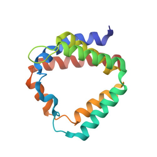
    The peridinin-chlorophyll a-protein (PCP) of dinoflagellates is unique among the large variety of natural photosynthetic light-harvesting systems. In contrast to other chlorophyll protein complexes, the soluble PCP is located in the thylakoid lumen, and the carotenoid pigments outnumber the chlorophylls. The structure of the PCP complex consists of two symmetric domains, each with a central chlorophyll a (Chl-a) surrounded by four peridinin molecules. The protein provides distinctive surroundings for the pigment molecules, and in PCP, the specific environment around each peridinin results in overlapping spectral line shapes, suggestive of different functions within the protein. One particular Per, Per-614, is hypothesized to show the strongest electronic interaction with the central Chl-a. We have performed an in vitro reconstitution of pigments into recombinant PCP apo-protein (RFPCP) and into a mutated protein with an altered environment near Per-614. Steady-state and transient optical spectroscopic experiments comparing the RFPCP complex with the reconstituted mutant protein identify specific amino acid-induced spectral shifts. The spectroscopic assignments are reinforced by a determination of the structures of both RFPCP and the mutant by x-ray crystallography to a resolution better than 1.5 A. RFPCP and mutated RFPCP are unique in representing crystal structures of in vitro reconstituted light-harvesting pigment-protein complexes.


  • Organizational Affiliation

    Biophysics, Department of Biology and Biotechnology, Ruhr-University Bochum, D-44780 Bochum, Germany.


Macromolecules
Find similar proteins by:  (by identity cutoff)  |  3D Structure
Entity ID: 1
MoleculeChains Sequence LengthOrganismDetailsImage
Peridinin-chlorophyll a-binding protein 1, chloroplasticA [auth M]151Amphidinium carteraeMutation(s): 3 
Gene Names: PCP
UniProt
Find proteins for P80484 (Amphidinium carterae)
Explore P80484 
Go to UniProtKB:  P80484
Entity Groups  
Sequence Clusters30% Identity50% Identity70% Identity90% Identity95% Identity100% Identity
UniProt GroupP80484
Sequence Annotations
Expand
  • Reference Sequence
Small Molecules
Ligands 5 Unique
IDChains Name / Formula / InChI Key2D Diagram3D Interactions
J7Z
Query on J7Z

Download Ideal Coordinates CCD File 
G [auth M](2S)-3-[(6-O-alpha-D-galactopyranosyl-beta-D-galactopyranosyl)oxy]-2-[(3Z,6Z,9Z,12Z,15Z)-octadeca-3,6,9,12,15-pentaenoyloxy]propyl (5Z,8Z,11Z,14Z,17Z)-icosa-5,8,11,14,17-pentaenoate
C53 H80 O15
GBUVPODJTFTLEC-ZHVHOYJFSA-N
CLA
Query on CLA

Download Ideal Coordinates CCD File 
B [auth M]CHLOROPHYLL A
C55 H72 Mg N4 O5
ATNHDLDRLWWWCB-AENOIHSZSA-M
PID
Query on PID

Download Ideal Coordinates CCD File 
C [auth M],
D [auth M],
E [auth M],
F [auth M]
PERIDININ
C39 H50 O7
UYRDHEJRPVSJFM-FROCQLDGSA-N
CD
Query on CD

Download Ideal Coordinates CCD File 
H [auth M],
I [auth M],
J [auth M],
K [auth M],
L [auth M]
CADMIUM ION
Cd
WLZRMCYVCSSEQC-UHFFFAOYSA-N
CL
Query on CL

Download Ideal Coordinates CCD File 
M,
N [auth M],
O [auth M],
P [auth M]
CHLORIDE ION
Cl
VEXZGXHMUGYJMC-UHFFFAOYSA-M
Experimental Data & Validation

Experimental Data

  • Method: X-RAY DIFFRACTION
  • Resolution: 1.45 Å
  • R-Value Free: 0.176 
  • R-Value Work: 0.155 
  • R-Value Observed: 0.156 
  • Space Group: C 2 2 21
Unit Cell:
Length ( Å )Angle ( ˚ )
a = 68.51α = 90
b = 81.73β = 90
c = 75.33γ = 90
Software Package:
Software NamePurpose
XDSdata scaling
MOLREPphasing
REFMACrefinement
XDSdata reduction
XSCALEdata scaling

Structure Validation

View Full Validation Report


Ligand Structure Quality Assessment 


Entry History 

Deposition Data

Revision History  (Full details and data files)

  • Version 1.0: 2009-11-24
    Type: Initial release
  • Version 1.1: 2011-07-13
    Changes: Version format compliance
  • Version 2.0: 2021-10-13
    Changes: Data collection, Database references, Derived calculations, Non-polymer description
  • Version 2.1: 2023-09-06
    Changes: Data collection, Refinement description