3IIQ

Crystallographic analysis of bacterial signal peptidase in ternary complex with Arylomycin A2 and a beta-sultam inhibitor


Experimental Data Snapshot

  • Method: X-RAY DIFFRACTION
  • Resolution: 2.00 Å
  • R-Value Free: 0.250 
  • R-Value Work: 0.207 
  • R-Value Observed: 0.210 

wwPDB Validation   3D Report Full Report


This is version 2.0 of the entry. See complete history


Literature

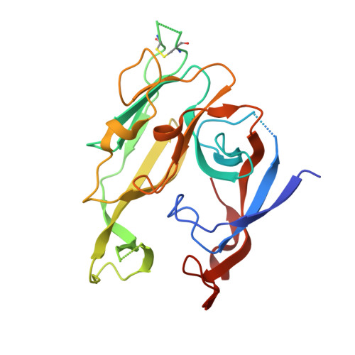
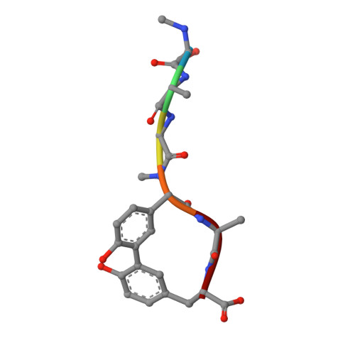
Crystallographic Analysis of Bacterial Signal Peptidase in Ternary Complex with Arylomycin A2 and a Beta-Sultam Inhibitor.

Luo, C.Roussel, P.Dreier, J.Page, M.G.Paetzel, M.

(2009) Biochemistry 48: 8976

  • DOI: https://doi.org/10.1021/bi9009538
  • Primary Citation of Related Structures:  
    3IIQ

  • PubMed Abstract: 

    Bacterial type I signal peptidase (SPase I), an essential membrane-bound endopeptidase with a unique Ser/Lys dyad mechanism, is being investigated as a potential novel antibiotic target. We present here binding and inhibition assays along with crystallographic data that shows that the lipohexapeptide-based natural product arylomycin A2 and the morpholino-beta-sultam derivative (BAL0019193) inhibit SPase I by binding to non-overlapping subsites near the catalytic center. The 2.0 A resolution crystal structure of the soluble catalytic domain of Escherichia coli SPase I (SPase I Delta2-75) in ternary complex with arylomycin A2 and BAL0019193 reveals the position of BAL0019193 adjacent to arylomycin A2 within the SPase I binding site. BAL0019193 binds in a noncovalent manner in close proximity to SPase I residues Ser88, Ser90, Lys145, Asn277, Ala279, and Glu307, as well as atom O45 of arylomycin A2. The binding mode of arylomycin A2 in this 2.0 A resolution ternary complex is compared to that seen in the previous 2.5 A resolution arylomycin A2-SPase cocrystal structure. This work contributes to our understanding of SPase I inhibitor/substrate recognition and should prove helpful in the further development of novel antibiotics based on the inhibition of SPase I.


  • Organizational Affiliation

    Department of Molecular Biology and Biochemistry, Simon Fraser University, South Science Building 8888 University Drive, Burnaby, British Columbia, V5A 1S6 Canada.


Macromolecules
Find similar proteins by:  (by identity cutoff)  |  3D Structure
Entity ID: 1
MoleculeChains Sequence LengthOrganismDetailsImage
SIGNAL PEPTIDASE I
A, B
249Escherichia coli K-12Mutation(s): 0 
Gene Names: LEPB
EC: 3.4.21.89
Membrane Entity: Yes 
UniProt
Find proteins for P00803 (Escherichia coli (strain K12))
Explore P00803 
Go to UniProtKB:  P00803
Entity Groups  
Sequence Clusters30% Identity50% Identity70% Identity90% Identity95% Identity100% Identity
UniProt GroupP00803
Sequence Annotations
Expand
  • Reference Sequence

Find similar proteins by:  Sequence   |   3D Structure  

Entity ID: 2
MoleculeChains Sequence LengthOrganismDetailsImage
ARYLOMYCIN A2
C, D
6Streptomyces sp.Mutation(s): 0 
Sequence Annotations
Expand
  • Reference Sequence
Small Molecules
Ligands 5 Unique
IDChains Name / Formula / InChI Key2D Diagram3D Interactions
TRT
Query on TRT

Download Ideal Coordinates CCD File 
F [auth A],
N [auth B]
FRAGMENT OF TRITON X-100
C21 H36 O4
HEUDUECKTWTQQR-UHFFFAOYSA-N
JZA
Query on JZA

Download Ideal Coordinates CCD File 
E [auth A],
M [auth B]
4-[(1,1-dioxido-1,2-thiazetidin-2-yl)carbonyl]morpholine
C7 H12 N2 O4 S
FLIYHWNBIOGIBH-UHFFFAOYSA-N
M12
Query on M12

Download Ideal Coordinates CCD File 
O [auth C],
P [auth D]
10-METHYLUNDECANOIC ACID
C12 H24 O2
QJRRBVNPIKYRQJ-UHFFFAOYSA-N
GOL
Query on GOL

Download Ideal Coordinates CCD File 
G [auth A],
H [auth A],
I [auth A],
J [auth A]
GLYCEROL
C3 H8 O3
PEDCQBHIVMGVHV-UHFFFAOYSA-N
CCN
Query on CCN

Download Ideal Coordinates CCD File 
K [auth A],
L [auth A]
ACETONITRILE
C2 H3 N
WEVYAHXRMPXWCK-UHFFFAOYSA-N
Modified Residues  1 Unique
IDChains TypeFormula2D DiagramParent
5PG
Query on 5PG
C, D
L-PEPTIDE LINKINGC9 H11 N O3GLY
Binding Affinity Annotations 
IDSourceBinding Affinity
JZA Binding MOAD:  3IIQ IC50: 6.10e+5 (nM) from 1 assay(s)
M12 Binding MOAD:  3IIQ IC50: 1.00e+6 (nM) from 1 assay(s)
Biologically Interesting Molecules (External Reference) 1 Unique
Experimental Data & Validation

Experimental Data

  • Method: X-RAY DIFFRACTION
  • Resolution: 2.00 Å
  • R-Value Free: 0.250 
  • R-Value Work: 0.207 
  • R-Value Observed: 0.210 
  • Space Group: P 43 21 2
Unit Cell:
Length ( Å )Angle ( ˚ )
a = 70.007α = 90
b = 70.007β = 90
c = 259.894γ = 90
Software Package:
Software NamePurpose
AMoREphasing
REFMACrefinement
HKL-2000data reduction
HKL-2000data scaling

Structure Validation

View Full Validation Report


Entry History 

Deposition Data

  • Released Date: 2009-09-01 
  • Deposition Author(s): Paetzel, M.

Revision History  (Full details and data files)

  • Version 1.0: 2009-09-01
    Type: Initial release
  • Version 1.1: 2011-06-14
    Changes: Version format compliance
  • Version 1.2: 2011-07-13
    Changes: Version format compliance
  • Version 1.3: 2011-07-27
    Changes: Advisory, Atomic model, Database references, Derived calculations, Non-polymer description, Structure summary
  • Version 1.4: 2012-12-12
    Changes: Other
  • Version 2.0: 2023-09-06
    Changes: Data collection, Database references, Derived calculations, Polymer sequence, Refinement description