3IFJ

Crystal structure of Mtu recA intein, splicing domain


Experimental Data Snapshot

  • Method: X-RAY DIFFRACTION
  • Resolution: 1.90 Å
  • R-Value Free: 0.260 
  • R-Value Work: 0.218 

wwPDB Validation   3D Report Full Report


This is version 2.0 of the entry. See complete history


Literature

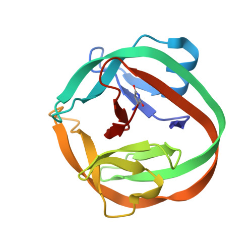
Selection and structure of hyperactive inteins: peripheral changes relayed to the catalytic center.

Hiraga, K.Soga, I.Dansereau, J.T.Pereira, B.Derbyshire, V.Du, Z.Wang, C.Van Roey, P.Belfort, G.Belfort, M.

(2009) J Mol Biol 393: 1106-1117

  • DOI: https://doi.org/10.1016/j.jmb.2009.08.074
  • Primary Citation of Related Structures:  
    3IFJ, 3IGD

  • PubMed Abstract: 

    Inteins are phylogenetically diverse self-splicing proteins that are of great functional, evolutionary, biotechnological, and medical interest. To address the relationship between intein structure and function, particularly with respect to regulating the splicing reaction, and to groom inteins for application, we developed a phage display system to extend current in vivo selection for enhanced intein function to selection in vitro. We thereby isolated inteins that can function under excursions in temperature, pH, and denaturing environment. Remarkably, most mutations mapped to the surface of the intein, remote from the active site. We chose two mutants with enhanced splicing activity for crystallography, one of which was also subjected to NMR analysis. These studies define a "ripple effect", whereby mutations in peripheral non-catalytic residues can cause subtle allosteric changes in the active-site environment in a way that facilitates intein activity. Altered salt-bridge formation and chemical shift changes of the mutant inteins provide a molecular rationale for their phenotypes. These fundamental insights will advance the utility of inteins in chemical biology, biotechnology, and medicine.


  • Organizational Affiliation

    Wadsworth Center, New York State Department of Health, 150 New Scotland Avenue, Albany, NY 12208, USA.


Macromolecules
Find similar proteins by:  (by identity cutoff)  |  3D Structure
Entity ID: 1
MoleculeChains Sequence LengthOrganismDetailsImage
Endonuclease PI-MtuI
A, B
139Mycobacterium tuberculosisMutation(s): 8 
Gene Names: MT2806MTV002.02crecARv2737c
EC: 3.1
UniProt
Find proteins for P9WHJ3 (Mycobacterium tuberculosis (strain ATCC 25618 / H37Rv))
Explore P9WHJ3 
Go to UniProtKB:  P9WHJ3
Entity Groups  
Sequence Clusters30% Identity50% Identity70% Identity90% Identity95% Identity100% Identity
UniProt GroupP9WHJ3
Sequence Annotations
Expand
  • Reference Sequence
Small Molecules
Modified Residues  1 Unique
IDChains TypeFormula2D DiagramParent
SNN
Query on SNN
A, B
L-PEPTIDE LINKINGC4 H6 N2 O2ASN
Experimental Data & Validation

Experimental Data

  • Method: X-RAY DIFFRACTION
  • Resolution: 1.90 Å
  • R-Value Free: 0.260 
  • R-Value Work: 0.218 
  • Space Group: P 21 21 21
Unit Cell:
Length ( Å )Angle ( ˚ )
a = 39.81α = 90
b = 86.56β = 90
c = 92.99γ = 90
Software Package:
Software NamePurpose
d*TREKdata scaling
PHASERphasing
CNSrefinement
PDB_EXTRACTdata extraction
CrystalCleardata collection
CrystalCleardata reduction
CrystalCleardata scaling

Structure Validation

View Full Validation Report


Entry History 

Deposition Data

Revision History  (Full details and data files)

  • Version 1.0: 2009-10-06
    Type: Initial release
  • Version 1.1: 2011-07-13
    Changes: Version format compliance
  • Version 1.2: 2017-08-16
    Changes: Refinement description, Source and taxonomy
  • Version 1.3: 2021-10-13
    Changes: Database references, Derived calculations
  • Version 1.4: 2023-09-06
    Changes: Data collection, Refinement description
  • Version 2.0: 2023-11-15
    Changes: Atomic model, Data collection, Derived calculations