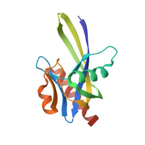
3I7U

Crystal structure of AP4A hydrolase (aq_158) from Aquifex aeolicus VF5


Experimental Data Snapshot

  • Method: X-RAY DIFFRACTION
  • Resolution: 1.80 Å
  • R-Value Free: 0.239 
  • R-Value Work: 0.203 
  • R-Value Observed: 0.203 

Starting Model: experimental
View more details

wwPDB Validation   3D Report Full Report


This is version 1.2 of the entry. See complete history


Literature

Free and ATP-bound structures of Ap(4)A hydrolase from Aquifex aeolicus V5

Jeyakanthan, J.Kanaujia, S.P.Nishida, Y.Nakagawa, N.Praveen, S.Shinkai, A.Kuramitsu, S.Yokoyama, S.Sekar, K.

(2010) Acta Crystallogr D Biol Crystallogr 66: 116-124

  • DOI: https://doi.org/10.1107/S0907444909047064
  • Primary Citation of Related Structures:  
    3I7U, 3I7V

  • PubMed Abstract: 

    Asymmetric diadenosine tetraphosphate (Ap(4)A) hydrolases degrade the metabolite Ap(4)A back into ATP and AMP. The three-dimensional crystal structure of Ap(4)A hydrolase (16 kDa) from Aquifex aeolicus has been determined in free and ATP-bound forms at 1.8 and 1.95 A resolution, respectively. The overall three-dimensional crystal structure of the enzyme shows an alphabetaalpha-sandwich architecture with a characteristic loop adjacent to the catalytic site of the protein molecule. The ATP molecule is bound in the primary active site and the adenine moiety of the nucleotide binds in a ring-stacking arrangement equivalent to that observed in the X-ray structure of Ap(4)A hydrolase from Caenorhabditis elegans. Binding of ATP in the active site induces local conformational changes which may have important implications in the mechanism of substrate recognition in this class of enzymes. Furthermore, two invariant water molecules have been identified and their possible structural and/or functional roles are discussed. In addition, modelling of the substrate molecule at the primary active site of the enzyme suggests a possible path for entry and/or exit of the substrate and/or product molecule.


  • Organizational Affiliation

    Life Science Group, National Synchrotron Radiation Research Center, 101 Hsin-Ann Road, Hsinchu Science Park, Hsinchu 30076, Taiwan.


Macromolecules
Find similar proteins by:  (by identity cutoff)  |  3D Structure
Entity ID: 1
MoleculeChains Sequence LengthOrganismDetailsImage
AP4A hydrolase
A, B, C, D
134Aquifex aeolicus VF5Mutation(s): 0 
Gene Names: apfAaq_158
UniProt
Find proteins for O66548 (Aquifex aeolicus (strain VF5))
Explore O66548 
Go to UniProtKB:  O66548
Entity Groups  
Sequence Clusters30% Identity50% Identity70% Identity90% Identity95% Identity100% Identity
UniProt GroupO66548
Sequence Annotations
Expand
  • Reference Sequence
Small Molecules
Ligands 6 Unique
IDChains Name / Formula / InChI Key2D Diagram3D Interactions
PG4
Query on PG4

Download Ideal Coordinates CCD File 
HA [auth D]TETRAETHYLENE GLYCOL
C8 H18 O5
UWHCKJMYHZGTIT-UHFFFAOYSA-N
PGE
Query on PGE

Download Ideal Coordinates CCD File 
FA [auth D],
GA [auth D]
TRIETHYLENE GLYCOL
C6 H14 O4
ZIBGPFATKBEMQZ-UHFFFAOYSA-N
TRS
Query on TRS

Download Ideal Coordinates CCD File 
R [auth C]2-AMINO-2-HYDROXYMETHYL-PROPANE-1,3-DIOL
C4 H12 N O3
LENZDBCJOHFCAS-UHFFFAOYSA-O
PEG
Query on PEG

Download Ideal Coordinates CCD File 
CA [auth D],
DA [auth D],
EA [auth D],
O [auth B]
DI(HYDROXYETHYL)ETHER
C4 H10 O3
MTHSVFCYNBDYFN-UHFFFAOYSA-N
EDO
Query on EDO

Download Ideal Coordinates CCD File 
AA [auth D]
BA [auth D]
H [auth A]
K [auth B]
L [auth B]
AA [auth D],
BA [auth D],
H [auth A],
K [auth B],
L [auth B],
M [auth B],
N [auth B],
W [auth D],
X [auth D],
Y [auth D],
Z [auth D]
1,2-ETHANEDIOL
C2 H6 O2
LYCAIKOWRPUZTN-UHFFFAOYSA-N
CL
Query on CL

Download Ideal Coordinates CCD File 
E [auth A]
F [auth A]
G [auth A]
I [auth B]
J [auth B]
E [auth A],
F [auth A],
G [auth A],
I [auth B],
J [auth B],
P [auth C],
Q [auth C],
S [auth D],
T [auth D],
U [auth D],
V [auth D]
CHLORIDE ION
Cl
VEXZGXHMUGYJMC-UHFFFAOYSA-M
Experimental Data & Validation

Experimental Data

  • Method: X-RAY DIFFRACTION
  • Resolution: 1.80 Å
  • R-Value Free: 0.239 
  • R-Value Work: 0.203 
  • R-Value Observed: 0.203 
  • Space Group: P 1 21 1
Unit Cell:
Length ( Å )Angle ( ˚ )
a = 85.589α = 90
b = 38.049β = 93.1
c = 87.592γ = 90
Software Package:
Software NamePurpose
HKL-2000data collection
MOLREPphasing
CNSrefinement
HKL-2000data reduction
HKL-2000data scaling

Structure Validation

View Full Validation Report


Entry History 

Deposition Data

Revision History  (Full details and data files)

  • Version 1.0: 2009-07-21
    Type: Initial release
  • Version 1.1: 2011-07-13
    Changes: Source and taxonomy, Version format compliance
  • Version 1.2: 2023-11-01
    Changes: Data collection, Database references, Derived calculations, Refinement description