3I4O | pdb_00003i4o

Crystal Structure of Translation Initiation Factor 1 from Mycobacterium tuberculosis


Experimental Data Snapshot

  • Method: X-RAY DIFFRACTION
  • Resolution: 1.47 Å
  • R-Value Free: 
    0.182 (Depositor), 0.183 (DCC) 
  • R-Value Work: 
    0.159 (Depositor), 0.163 (DCC) 
  • R-Value Observed: 
    0.160 (Depositor) 

wwPDB Validation 3D Report Full Report

Validation slider image for 3I4O

This is version 1.4 of the entry. See complete history

Literature

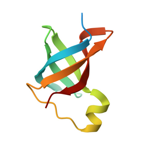
Structure of translation initiation factor 1 from Mycobacterium tuberculosis and inferred binding to the 30S ribosomal subunit.

Hatzopoulos, G.N.Mueller-Dieckmann, J.

(2010) FEBS Lett 584: 1011-1015

  • DOI: https://doi.org/10.1016/j.febslet.2010.01.051
  • Primary Citation Related Structures: 
    3I4O

  • PubMed Abstract: 

    The crystal structure of the free form of IF1 from Mycobacterium tuberculosis has been determined at 1.47 A resolution. The structure adopts the expected OB fold and matches the high structural conservation among IF1 orthologues. In order to further explore the function of Mtb-IF1, we built a model of its interaction with the 30S ribosomal subunit based on the crystal structure of the complex from Thermus thermophilus. The model suggests that several functionally important side chain residues undergo large movements while the rest of the protein in complex shows only very limited conformational change as compared to its form in solution.


  • Organizational Affiliation
    • EMBL Hamburg Outstation, c/o DESY, Notkestr. 85, D-22603 Hamburg, Germany.

Macromolecule Content 

  • Total Structure Weight: 18.27 kDa 
  • Atom Count: 1,418 
  • Modeled Residue Count: 135 
  • Deposited Residue Count: 158 
  • Unique protein chains: 1

Macromolecules

Find similar proteins by:|  3D Structure
Entity ID: 1
MoleculeChains  Sequence LengthOrganismDetailsImage
Translation initiation factor IF-1
A, B
79Mycobacterium tuberculosis H37RvMutation(s): 0 
Gene Names: infAMCB1222.32cMT3568MTCY13E12.15cRv3462cRv3462c (infA)
UniProt
Find proteins for P9WKK3 (Mycobacterium tuberculosis (strain ATCC 25618 / H37Rv))
Explore P9WKK3 
Go to UniProtKB:  P9WKK3
Entity Groups
Sequence Clusters30% Identity50% Identity70% Identity90% Identity95% Identity100% Identity
UniProt GroupP9WKK3
Sequence Annotations
Expand
Reference Sequence

Experimental Data & Validation

Experimental Data

  • Method: X-RAY DIFFRACTION
  • Resolution: 1.47 Å
  • R-Value Free:  0.182 (Depositor), 0.183 (DCC) 
  • R-Value Work:  0.159 (Depositor), 0.163 (DCC) 
  • R-Value Observed: 0.160 (Depositor) 
Space Group: P 21 21 2
Unit Cell:
Length ( Å )Angle ( ˚ )
a = 73.95α = 90
b = 76.54β = 90
c = 28.03γ = 90
Software Package:
Software NamePurpose
DENZOdata reduction
SCALEPACKdata scaling
SHELXphasing
REFMACrefinement
PDB_EXTRACTdata extraction
BESTdata collection
SHELXDphasing

Structure Validation

View Full Validation Report


Entry History 

Deposition Data

Revision History  (Full details and data files)

  • Version 1.0: 2010-02-23
    Type: Initial release
  • Version 1.1: 2011-07-13
    Changes: Version format compliance
  • Version 1.2: 2013-11-13
    Changes: Database references
  • Version 1.3: 2017-11-01
    Changes: Refinement description
  • Version 1.4: 2024-03-20
    Changes: Data collection, Database references