3HZB

Crystal structure of a betagamma-crystallin domain from Flavobacterium johnsoniae


Experimental Data Snapshot

  • Method: X-RAY DIFFRACTION
  • Resolution: 1.74 Å
  • R-Value Free: 0.223 
  • R-Value Work: 0.192 

wwPDB Validation   3D Report Full Report


This is version 1.3 of the entry. See complete history


Literature

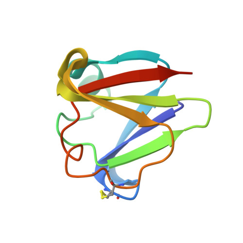
The betagamma-crystallin superfamily contains a universal motif for binding calcium

Aravind, P.Mishra, A.Suman, S.K.Jobby, M.K.Sankaranarayanan, R.Sharma, Y.

(2009) Biochemistry 48: 12180-12190

  • DOI: https://doi.org/10.1021/bi9017076
  • Primary Citation of Related Structures:  
    3HZ2, 3HZB, 3I9H, 3IAJ

  • PubMed Abstract: 

    The betagamma-crystallin superfamily consists of evolutionarily related proteins with domain topology similar to lens beta- and gamma-crystallins, formed from duplicated Greek key motifs. Ca(2+) binding was found in a few betagamma-crystallin members earlier, although its prevalence and diversity as inherent molecular properties among members of the superfamily are not well studied. To increase our understanding of Ca(2+) binding in various betagamma-crystallins, we undertook comprehensive structural and Ca(2+)-binding studies of seven members of the superfamily from bacteria, archaea, and vertebrates, including determination of high-resolution crystal structures of three proteins. Our structural observations show that the determinants of Ca(2+) coordination remain conserved in the form of an N/D-N/D-#-I-S/T-S motif in all domains. However, binding of Ca(2+) elicits varied physicochemical responses, ranging from passive sequestration to active stabilization. The motif in this superfamily is modified in some members like lens crystallins where Ca(2+)-binding abilities are partly or completely compromised. We show that reduction or loss of Ca(2+) binding in members of the superfamily, particularly in vertebrates, is due to the selective presence of unfavorable amino acids (largely Arg) at key Ca(2+)-ligation positions and that engineering of the canonical Ca(2+)-binding residues can confer binding activity on an otherwise inactive domain. Through this work, we demonstrate that betagamma-crystallins with the N/D-N/D-#-I-S/T-S motif form an extensive set of Ca(2+)-binding proteins prevalent in all of the three kingdoms of life.


  • Organizational Affiliation

    Centre for Cellular and Molecular Biology, Council of Scientific and Industrial Research, Uppal Road, Hyderabad 500007, India.


Macromolecules
Find similar proteins by:  (by identity cutoff)  |  3D Structure
Entity ID: 1
MoleculeChains Sequence LengthOrganismDetailsImage
Carbohydrate binding protein
A, B, C, D, E
A, B, C, D, E, F, G, H
90Flavobacterium johnsoniae UW101Mutation(s): 0 
Gene Names: 5093804
UniProt
Find proteins for A5FC88 (Flavobacterium johnsoniae (strain ATCC 17061 / DSM 2064 / JCM 8514 / NBRC 14942 / NCIMB 11054 / UW101))
Explore A5FC88 
Go to UniProtKB:  A5FC88
Entity Groups  
Sequence Clusters30% Identity50% Identity70% Identity90% Identity95% Identity100% Identity
UniProt GroupA5FC88
Sequence Annotations
Expand
  • Reference Sequence
Small Molecules
Ligands 1 Unique
IDChains Name / Formula / InChI Key2D Diagram3D Interactions
CA
Query on CA

Download Ideal Coordinates CCD File 
AA [auth H]
BA [auth H]
I [auth A]
J [auth A]
K [auth A]
AA [auth H],
BA [auth H],
I [auth A],
J [auth A],
K [auth A],
L [auth B],
M [auth B],
N [auth C],
O [auth C],
P [auth C],
Q [auth D],
R [auth D],
S [auth E],
T [auth E],
U [auth E],
V [auth F],
W [auth F],
X [auth G],
Y [auth G],
Z [auth G]
CALCIUM ION
Ca
BHPQYMZQTOCNFJ-UHFFFAOYSA-N
Experimental Data & Validation

Experimental Data

  • Method: X-RAY DIFFRACTION
  • Resolution: 1.74 Å
  • R-Value Free: 0.223 
  • R-Value Work: 0.192 
  • Space Group: P 1 2 1
Unit Cell:
Length ( Å )Angle ( ˚ )
a = 53.647α = 90
b = 99.279β = 89.99
c = 64.101γ = 90
Software Package:
Software NamePurpose
DENZOdata reduction
SCALEPACKdata scaling
MOLREPphasing
CNSrefinement
PDB_EXTRACTdata extraction

Structure Validation

View Full Validation Report


Entry History 

Deposition Data

Revision History  (Full details and data files)

  • Version 1.0: 2009-12-01
    Type: Initial release
  • Version 1.1: 2011-07-13
    Changes: Version format compliance
  • Version 1.2: 2017-11-01
    Changes: Refinement description
  • Version 1.3: 2023-11-01
    Changes: Data collection, Database references, Derived calculations, Refinement description