3HQA

Crystal structure of human desarg-C5A


Experimental Data Snapshot

  • Method: X-RAY DIFFRACTION
  • Resolution: 2.59 Å
  • R-Value Free: 0.253 
  • R-Value Work: 0.213 
  • R-Value Observed: 0.215 

wwPDB Validation   3D Report Full Report


This is version 1.5 of the entry. See complete history


Literature

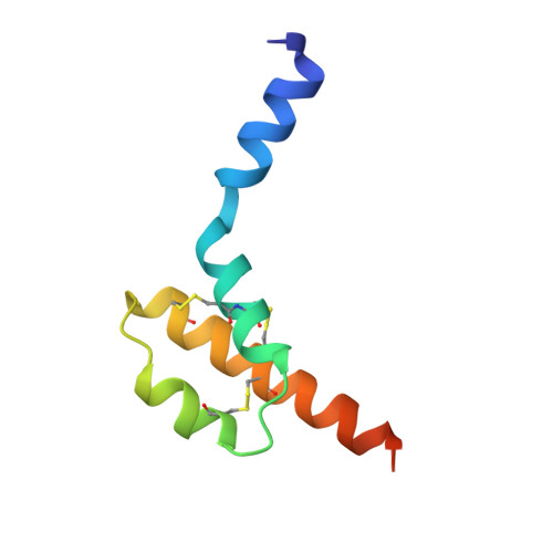
Structure of human desArg-C5a.

Cook, W.J.Galakatos, N.Boyar, W.C.Walter, R.L.Ealick, S.E.

(2010) Acta Crystallogr D Biol Crystallogr 66: 190-197

  • DOI: https://doi.org/10.1107/S0907444909049051
  • Primary Citation of Related Structures:  
    3HQA, 3HQB

  • PubMed Abstract: 

    The anaphylatoxin C5a is derived from the complement component C5 during activation of the complement cascade. It is an important component in the pathogenesis of a number of inflammatory diseases. NMR structures of human and porcine C5a have been reported; these revealed a four-helix bundle stabilized by three disulfide bonds. The crystal structure of human desArg-C5a has now been determined in two crystal forms. Surprisingly, the protein crystallizes as a dimer and each monomer in the dimer has a three-helix core instead of the four-helix bundle noted in the NMR structure determinations. Furthermore, the N-terminal helices of the two monomers occupy different positions relative to the three-helix core and are completely different from the NMR structures. The physiological significance of these structural differences is unknown.


  • Organizational Affiliation

    University of Alabama at Birmingham, Birmingham, AL 35294, USA. wjcook@uab.edu


Macromolecules
Find similar proteins by:  (by identity cutoff)  |  3D Structure
Entity ID: 1
MoleculeChains Sequence LengthOrganismDetailsImage
Complement C5
A, B
73Homo sapiensMutation(s): 0 
Gene Names: C5CPAMD4
UniProt & NIH Common Fund Data Resources
Find proteins for P01031 (Homo sapiens)
Explore P01031 
Go to UniProtKB:  P01031
PHAROS:  P01031
GTEx:  ENSG00000106804 
Entity Groups  
Sequence Clusters30% Identity50% Identity70% Identity90% Identity95% Identity100% Identity
UniProt GroupP01031
Sequence Annotations
Expand
  • Reference Sequence
Experimental Data & Validation

Experimental Data

  • Method: X-RAY DIFFRACTION
  • Resolution: 2.59 Å
  • R-Value Free: 0.253 
  • R-Value Work: 0.213 
  • R-Value Observed: 0.215 
  • Space Group: P 41 21 2
Unit Cell:
Length ( Å )Angle ( ˚ )
a = 50.61α = 90
b = 50.61β = 90
c = 117.49γ = 90
Software Package:
Software NamePurpose
PHENIXrefinement
PHASERphasing
CNSrefinement
X-GENdata reduction
X-GENdata scaling

Structure Validation

View Full Validation Report


Entry History 

Deposition Data

Revision History  (Full details and data files)

  • Version 1.0: 2010-02-02
    Type: Initial release
  • Version 1.1: 2011-07-13
    Changes: Version format compliance
  • Version 1.2: 2012-03-28
    Changes: Database references
  • Version 1.3: 2017-11-01
    Changes: Refinement description
  • Version 1.4: 2019-07-24
    Changes: Data collection, Refinement description
  • Version 1.5: 2023-09-06
    Changes: Data collection, Database references, Refinement description