3HP8 | pdb_00003hp8

Crystal structure of a designed Cyanovirin-N homolog lectin; LKAMG, bound to sucrose


Experimental Data Snapshot

  • Method: X-RAY DIFFRACTION
  • Resolution: 2.00 Å
  • R-Value Free: 
    0.277 (Depositor), 0.280 (DCC) 
  • R-Value Work: 
    0.224 (Depositor), 0.226 (DCC) 
  • R-Value Observed: 
    0.227 (Depositor) 

Starting Model: experimental
View more details

wwPDB Validation 3D Report Full Report

Validation slider image for 3HP8

This is version 2.1 of the entry. See complete history

Literature

A designed chimeric cyanovirin-N homolog lectin: Structure and molecular basis of sucrose binding.

Koharudin, L.M.Furey, W.Gronenborn, A.M.

(2009) Proteins 77: 904-915

  • DOI: https://doi.org/10.1002/prot.22514
  • Primary Citation Related Structures: 
    3HNU, 3HNX, 3HP8

  • PubMed Abstract: 

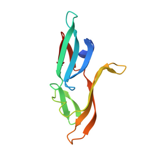
    The NMR and X-ray structures of a designed chimeric cyanovirin-N homolog (CVNH) protein were determined. The individual halves of the structure are similar to their counterparts in the parent proteins, with domains A and B resembling the structures of TbCVNH and NcCVNH, respectively. No significant differences between the solution and crystal conformations were observed, although details in loop conformations and distinct crystal packing-induced features are present. Carbohydrate binding studies by NMR revealed affinity and specificity for Glc alpha(1-2)Frc and Man alpha(1-2)Man, and the parental half that is devoid of any sucrose affinity in NcCVNH was transformed into a genuine sucrose binding site in the context of the chimera. The atomic details of sugar recognition are seen in the crystal structure of the protein with two bound Glc alpha(1-2)Frc molecules. Both sugars exhibit different conformations around the glycosidic bond and engage in unique hydrogen bonding networks in the two sites. Although the protein is able to bind two Man alpha(1-2)Man molecules, a property associated with HIV-inactivation, no anti-HIV activity was observed for the hybrid protein. These results provide the structural basis for sugar recognition in the CVNH family and aid in deciphering the relationship between sugar binding and anti-HIV activity.


  • Organizational Affiliation
    • Department of Structural Biology, University of Pittsburgh School of Medicine, Pittsburgh, Pennsylvania 15260, USA.

Macromolecule Content 

  • Total Structure Weight: 26.45 kDa 
  • Atom Count: 2,067 
  • Modeled Residue Count: 214 
  • Deposited Residue Count: 220 
  • Unique protein chains: 1

Macromolecules

Find similar proteins by:|  3D Structure
Entity ID: 1
MoleculeChains  Sequence LengthOrganismDetailsImage
Cyanovirin-N-like protein
A, B
110Tuber borchiiNeurospora crassaMutation(s): 0 
UniProt
Find proteins for Q5MK11 (Tuber borchii)
Explore Q5MK11 
Go to UniProtKB:  Q5MK11
Find proteins for Q7S6U4 (Neurospora crassa (strain ATCC 24698 / 74-OR23-1A / CBS 708.71 / DSM 1257 / FGSC 987))
Explore Q7S6U4 
Go to UniProtKB:  Q7S6U4
Entity Groups
Sequence Clusters30% Identity50% Identity70% Identity90% Identity95% Identity100% Identity
UniProt GroupsQ5MK11Q7S6U4
Sequence Annotations
Expand
Reference Sequence

Oligosaccharides

Help  
Entity ID: 2
MoleculeChains Length2D Diagram GlycosylationD Interactions
beta-D-fructofuranose-(2-1)-alpha-D-glucopyranose
C, D, E, F
2N/A
Glycosylation Resources
GlyTouCan: G05551OP
GlyCosmos: G05551OP

Biologically Interesting Molecules (External Reference) 

1 Unique

Experimental Data & Validation

Experimental Data

  • Method: X-RAY DIFFRACTION
  • Resolution: 2.00 Å
  • R-Value Free:  0.277 (Depositor), 0.280 (DCC) 
  • R-Value Work:  0.224 (Depositor), 0.226 (DCC) 
  • R-Value Observed: 0.227 (Depositor) 
Space Group: P 1 21 1
Unit Cell:
Length ( Å )Angle ( ˚ )
a = 44.497α = 90
b = 39.298β = 97.64
c = 86.134γ = 90
Software Package:
Software NamePurpose
StructureStudiodata collection
PHASERphasing
REFMACrefinement
d*TREKdata reduction
d*TREKdata scaling

Structure Validation

View Full Validation Report


Entry History 

Deposition Data

Revision History  (Full details and data files)

  • Version 1.0: 2009-06-23
    Type: Initial release
  • Version 1.1: 2011-07-13
    Changes: Version format compliance
  • Version 1.2: 2017-08-16
    Changes: Refinement description, Source and taxonomy
  • Version 2.0: 2020-07-29
    Type: Remediation
    Reason: Carbohydrate remediation
    Changes: Advisory, Atomic model, Data collection, Database references, Derived calculations, Non-polymer description, Structure summary
  • Version 2.1: 2023-09-06
    Changes: Data collection, Database references, Refinement description, Structure summary