Discovery of N-substituted pyridinones as potent and selective inhibitors of p38 kinase.
Selness, S.R., Devraj, R.V., Monahan, J.B., Boehm, T.L., Walker, J.K., Devadas, B., Durley, R.C., Kurumbail, R., Shieh, H., Xing, L., Hepperle, M., Rucker, P.V., Jerome, K.D., Benson, A.G., Marrufo, L.D., Madsen, H.M., Hitchcock, J., Owen, T.J., Christie, L., Promo, M.A., Hickory, B.S., Alvira, E., Naing, W., Blevis-Bal, R.(2009) Bioorg Med Chem Lett 19: 5851-5856
- PubMed: 19751974 Search on PubMed
- DOI: https://doi.org/10.1016/j.bmcl.2009.08.082
- Primary Citation Related Structures:
3HP2, 3HP5 - PubMed Abstract:
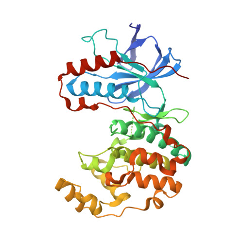
The identification and evolution of a series of potent and selective p38 inhibitors is described. p38 inhibitors based on a N-benzyl pyridinone high-throughput screening hit were prepared and their SAR explored. Their design was guided by ligand bound co-crystals of p38alpha. These efforts resulted in the identification of 12r and 19 as orally active inhibitors of p38 with significant efficacy in both acute and chronic models of inflammation.
- Department of Medicinal Chemistry, Pfizer Corporation, 700 Chesterfield Parkway West, Chesterfield, MO 63017, USA. shaun.r.selness@pfizer.com
Organizational Affiliation: